 Back
BackMini-Textbook Study Guide: Central Nervous System Structure & Function
Study Guide - Smart Notes
 Tailored notes based on your materials, expanded with key definitions, examples, and context.
Basic Embryology of the Central Nervous System
Developmental Landmarks and Brain Vesicles
The central nervous system (CNS) develops from the neural tube, which forms primary and secondary brain vesicles. These vesicles give rise to the major regions of the adult brain and spinal cord. - Primary brain vesicles: Prosencephalon (forebrain), Mesencephalon (midbrain), Rhombencephalon (hindbrain) - Secondary brain vesicles: Telencephalon, Diencephalon, Mesencephalon, Metencephalon, Myelencephalon - Adult brain structures: Cerebrum, diencephalon, brain stem (midbrain, pons, medulla oblongata), cerebellum, spinal cord - Neural canal regions: Lateral ventricles, third ventricle, cerebral aqueduct, fourth ventricle, central canal 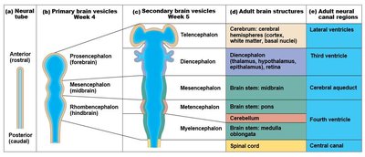
Embryonic Growth and Flexures
During embryonic development, the brain undergoes rapid growth, forming flexures and folds that shape its final structure. - Flexures: Bending of the neural tube creates major divisions and landmarks - Folding: Surface of the cerebrum begins to fold, forming gyri and sulci - Enclosure: Cerebral hemispheres grow to enclose the diencephalon and brain stem 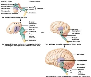
Gross Anatomy of the Brain
Gyri, Sulci, and Fissures
The surface of the cerebrum is highly folded, increasing surface area for cortical processing. - Gyrus: A ridge or fold on the cerebral surface - Sulcus: A shallow groove between gyri - Fissure: A deep groove separating major brain regions - Formation: These landmarks form embryonically and are associated with specific functions 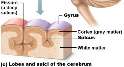
Lobes and Major Landmarks
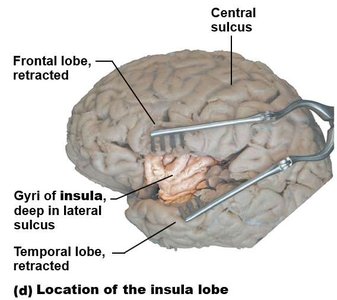
The cerebrum is divided into lobes by prominent sulci and fissures. - Frontal lobe: Responsible for motor function, problem solving, and speech - Parietal lobe: Processes sensory information - Occipital lobe: Visual processing center - Temporal lobe: Auditory processing and memory - Insula: Deep to the lateral sulcus, involved in taste and visceral sensation - Central sulcus: Separates frontal and parietal lobes - Lateral sulcus: Separates temporal lobe from frontal and parietal lobes - Parieto-occipital sulcus: Separates parietal and occipital lobes - Transverse cerebral fissure: Separates cerebrum from cerebellum 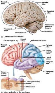
Ventricles of the Brain
Structure and Function
The brain contains fluid-filled chambers called ventricles, which are continuous with the central canal of the spinal cord. - Lateral ventricles: Located in each cerebral hemisphere - Third ventricle: Located in the diencephalon - Cerebral aqueduct: Connects third and fourth ventricles - Fourth ventricle: Located between pons and cerebellum; connects to subarachnoid space via apertures - Function: Circulation of cerebrospinal fluid (CSF) 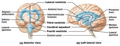
Cerebral Hemispheres and Functional Areas
Cerebral Regions
The cerebrum consists of three main regions: cortex, white matter, and cerebral nuclei. - Cortex: Gray matter responsible for higher-order functions - White matter: Communication tracts between regions - Cerebral nuclei: Basal nuclei regulate voluntary motor activity 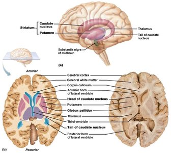
Functional Areas of the Cortex
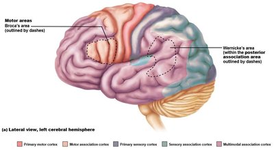
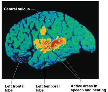
The cortex is organized into distinct functional areas. - Sensory areas: Conscious awareness of stimuli - Sensory association areas: Integration and interpretation of sensory input - Motor areas: Control voluntary muscle contraction - Motor association areas: Integrate sensory information to form motor plans - Multimodal association areas: Integrate multiple sensory inputs for complex understanding 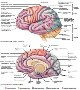
Cerebral Tracts
Types of Fibers
White matter tracts facilitate communication within and between brain regions. - Commissural fibers: Connect corresponding regions of the two hemispheres (e.g., corpus callosum) - Association fibers: Connect areas within a single hemisphere - Projection fibers: Connect the cerebrum with lower parts of the CNS 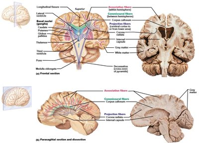
Diencephalon
Thalamus
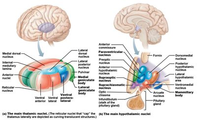
The thalamus is a major relay station for sensory and motor information. - Location: Surrounds the third ventricle - Function: Processes and modifies information passing to/from the cortex - Interthalamic adhesion: Joins the two halves of the thalamus 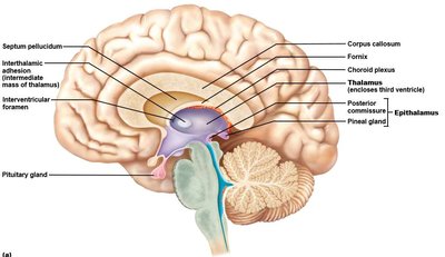
Hypothalamus
The hypothalamus is the visceral control center of the body. - Location: Inferolateral walls of the third ventricle, between optic chiasma and mammillary bodies - Function: Regulates autonomic functions, endocrine activity, and homeostasis - Infundibulum: Connects hypothalamus to the posterior pituitary gland
Brain Stem
Midbrain
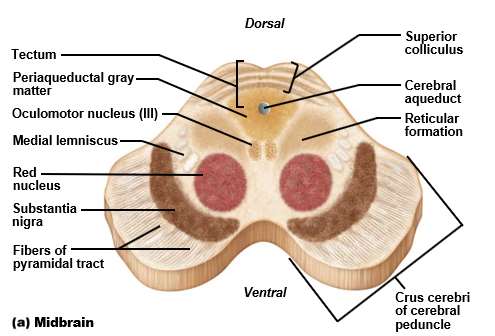
The midbrain connects the diencephalon to the pons and contains important motor and sensory pathways. - Cerebral aqueduct: Central cavity of the midbrain - Tectum: Posterior region containing colliculi - Cerebral peduncles: Anterior region containing pyramidal motor tracts - Cranial nerves: Origin of III (oculomotor) and IV (trochlear) 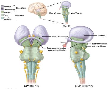
Pons
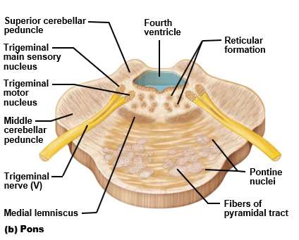
The pons is a relay center between the cerebrum and cerebellum and contains nuclei for cranial nerves. - Location: Separated from cerebellum by fourth ventricle - Pontine nuclei: Relay information to cerebellum via cerebellar peduncles - Cranial nerves: Origin of V (trigeminal), VI (abducens), VII (facial) 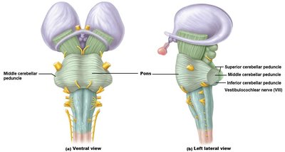
Medulla Oblongata
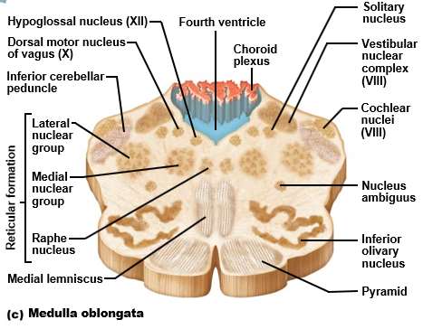
The medulla oblongata controls vital autonomic functions and is the site of decussation of pyramidal tracts. - Contains: Fourth ventricle, choroid plexus, pyramids - Decussation: Crossing over of motor output tracts - Cranial nerves: Origin of VIII (vestibulocochlear), IX (glossopharyngeal), X (vagus), XI (accessory), XII (hypoglossal) 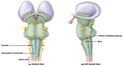
Cerebellum
Structure and Function
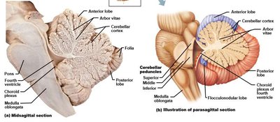
The cerebellum coordinates smooth, skilled voluntary movements and maintains balance. - Location: Dorsal to pons and medulla oblongata, separated by fourth ventricle - Hemispheres: Connected by vermis, divided into anterior, posterior, and flocculonodular lobes - Cortex: Gray matter - Arbor vitae: Tracts of white matter connecting to cerebellar peduncles 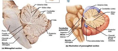
Spinal Cord
Gross Anatomy
The spinal cord is protected by meninges and the vertebral column, and extends from the foramen magnum to L1/L2. - Conus medullaris: Inferior end of the spinal cord - Filum terminale: Filament anchoring the cord to the coccyx - Cauda equina: Collection of spinal nerve roots below L2 - Cervical and lumbar enlargements: Regions with increased neural input/output
External and Internal Anatomy
- Dorsal root ganglia: Used to determine orientation and anatomical position - Spinal nerves: Formed by merging dorsal and ventral roots; named for vertebrae where they emerge - Gray matter: Divided into horns (cell bodies of processing or motor neurons) - White matter: Divided into tracts (funiculi): dorsal, lateral, ventral
Ascending and Descending Tracts
- Ascending tracts: 3-neuron pathway carrying sensory information up to the brain (e.g., spinocerebellar, dorsal column-medial lemniscus, spinothalamic) - Descending tracts: 2-neuron pathway carrying motor information down from the brain (e.g., pyramidal/corticospinal, rubrospinal) Equation for ascending pathway: Equation for descending pathway:
Cranial Nerves
Overview and Mnemonics
There are twelve pairs of cranial nerves, each with specific functions and origins. - Mnemonic for names: Oh, Once, One, Takes, The, Anatomy, Final, Very, Good, Vacations, Are, Happening - Mnemonic for function: Some Say Money Matters But My Brother Says Big Brains Matter More - Origin: Temporal lobe, occipital lobe, brain stem (midbrain, pons, medulla oblongata)
# | Name | Mnemonic | Function Mnemonic | Origin |
|---|---|---|---|---|
I | Olfactory | Oh | Some | Temporal lobe |
II | Optic | Once | Say | Occipital lobe |
III | Oculomotor | One | Marry | Midbrain |
IV | Trochlear | Takes | Money | Midbrain |
V | Trigeminal | The | But | Pons |
VI | Abducens | Anatomy | My | Pons |
VII | Facial | Final | Brother | Pons |
VIII | Vestibulocochlear | Very | Says | Medulla oblongata |
IX | Glossopharyngeal | Good | Bad | Medulla oblongata |
X | Vagus | Vacations | Business | Medulla oblongata |
XI | Accessory | Are | Marry | Medulla oblongata |
XII | Hypoglossal | Happening | Money | Medulla oblongata |
Example: The trigeminal nerve (V) originates from the pons and is responsible for facial sensation and chewing movements. Additional info: Cranial nerves are often tested in clinical exams to assess neurological function. ----------------------------------------
