Back
BackMini-Textbook Study Guide: Histology and Cell Transport for ANP College
Study Guide - Smart Notes
 Tailored notes based on your materials, expanded with key definitions, examples, and context.
Cell Transport Mechanisms
Passive and Active Transport
Cells utilize various mechanisms to move substances across their membranes. Passive transport does not require energy, while active transport does. Understanding these processes is essential for grasping cellular physiology.
Simple Diffusion: Movement of molecules from high to low concentration without assistance.
Osmosis: Diffusion of water across a selectively permeable membrane.
Facilitated Diffusion: Movement of molecules via protein channels or carriers.
Phagocytosis: An active transport process where cells engulf large particles (not passive).
Example: Water moving from an area of low solute concentration to high solute concentration across a membrane is osmosis.
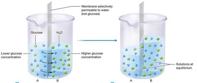
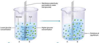
Osmosis Illustrated
Osmosis is a specific type of passive transport where water moves across a membrane that is permeable to water but not to solutes like glucose. This process continues until equilibrium is reached.
Key Point: Water moves from the side with lower solute concentration to the side with higher solute concentration.
Application: Osmosis is critical in maintaining cell volume and fluid balance in tissues.
Organelle Functions
Cell organelles perform specialized functions. The rough endoplasmic reticulum (RER) is responsible for producing and folding proteins, while other organelles have distinct roles.
Mitochondria: Generate ATP through cellular respiration.
Rough Endoplasmic Reticulum: Synthesizes and folds proteins; studded with ribosomes.
Smooth Endoplasmic Reticulum: Synthesizes lipids and detoxifies chemicals.
Golgi Apparatus: Modifies, sorts, and packages proteins for secretion.
Example: Cells with abundant RER are typically involved in protein secretion, such as glandular cells.
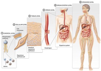
Levels of Structural Organization
Hierarchy of Biological Organization
Biological systems are organized into hierarchical levels, each with increasing complexity. This organization is fundamental to understanding anatomy and physiology.
Chemical Level: Atoms and molecules (e.g., phospholipids).
Cellular Level: Cells and their organelles (e.g., epithelial cells).
Tissue Level: Groups of similar cells performing a common function (e.g., stratified squamous epithelium).
Organ Level: Structures composed of multiple tissue types (e.g., esophagus).
Organ System Level: Groups of organs working together (e.g., digestive system).
Organism Level: The complete living being.
Histology: The Study of Tissues
Definition and Major Types of Tissues
Histology is the study of tissues, which are groups of structurally and functionally related cells and their external environment. There are four major tissue types:
Epithelial Tissue: Sheets of tightly packed cells with little ECM; lines surfaces and cavities.
Connective Tissue: Connects, supports, and protects other tissues; abundant ECM.
Muscle Tissue: Cells that contract to generate force; little ECM.
Nervous Tissue: Cells that generate and transmit electrical signals; support cells included.
Additional info: Organs are composed of two or more tissue types working together.
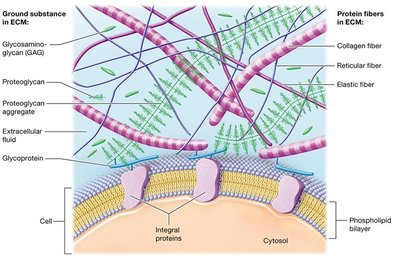
The Extracellular Matrix (ECM)
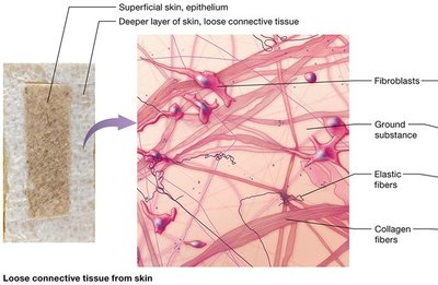
The extracellular matrix is the material surrounding cells in a tissue. It provides structural support, resists mechanical forces, and holds cells in place.
Ground Substance: Gel-like material containing water, ions, nutrients, and solutes.
Protein Fibers: Collagen (tensile strength), elastic (stretch and recoil), reticular (meshwork support).
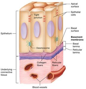
Cell Junctions
Cell junctions are specialized structures that link neighboring cells, contributing to tissue integrity and communication.
Tight Junctions: Seal spaces between cells, making them impermeable to macromolecules.
Desmosomes: Provide mechanical strength by anchoring cells together.
Gap Junctions: Allow passage of small molecules and electrical signals between cells.

Epithelial Tissue
Structure and Function
Epithelial tissues cover internal and external surfaces, acting as barriers and performing various functions.
Protection: Shields underlying tissues.
Immune Defense: Blocks pathogens.
Secretion: Forms glands that release substances.
Transport: Selectively allows substances to cross.
Sensation: Contains sensory nerves.
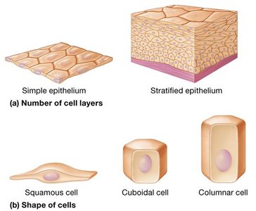
Classification of Epithelial Cells
Epithelial tissues are classified by the number of cell layers and the shape of the cells.
Simple Epithelium: Single layer of cells.
Stratified Epithelium: Multiple layers of cells.
Squamous Cells: Flat and thin.
Cuboidal Cells: Cube-shaped.
Columnar Cells: Tall and column-like.
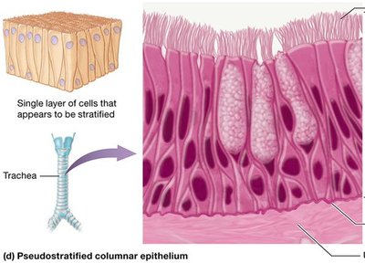
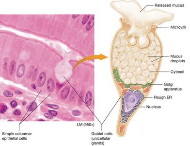
Pseudostratified Epithelium
Pseudostratified epithelium appears to have multiple layers but is actually a single layer of columnar cells, often containing goblet cells that secrete mucus.
Location: Commonly found in the trachea.
Function: Mucus secretion and trapping particles.
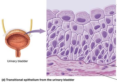
Transitional Epithelium
Transitional epithelium is unique to the urinary bladder. Its cells change shape depending on bladder fullness, appearing squamous when stretched and cuboidal when relaxed.
Function: Allows bladder to expand and contract.
Glandular Epithelium
Glands are structures that secrete products. They arise from epithelial tissue and are classified as exocrine or endocrine.
Exocrine Glands: Secrete products via ducts to surfaces; local action.
Endocrine Glands: Secrete hormones directly into the blood; systemic action.
Unicellular Exocrine Glands: Goblet Cells
Goblet cells are the most common unicellular exocrine glands, secreting mucus in various epithelial tissues.
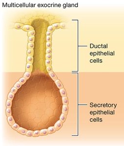
Multicellular Exocrine Glands
Multicellular exocrine glands consist of ductal and secretory epithelial cells, forming complex structures for secretion.
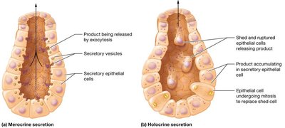
Exocrine Secretion Types
Exocrine glands release their products by different mechanisms:
Merocrine Secretion: Products released by exocytosis (e.g., salivary glands).
Holocrine Secretion: Products released when cells rupture and die (e.g., sebaceous glands).
Connective Tissue
Functions and Types
Connective tissue binds, supports, protects, and transports substances. It is divided into connective tissue proper and specialized connective tissue.
Connective Tissue Proper: Loose, dense, reticular, and adipose tissues.
Specialized Connective Tissue: Cartilage, bone, and blood.
Loose Connective Tissue
Loose connective tissue contains abundant ground substance, all three protein fibers, and various cells including fibroblasts.
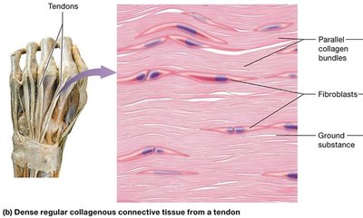
Dense Connective Tissue
Dense connective tissue is primarily composed of protein fibers, which may be arranged in parallel bundles or irregularly.
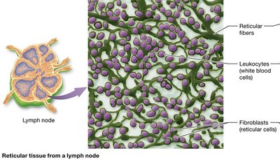
Reticular Tissue
Reticular tissue contains numerous reticular fibers, supporting small structures and housing white blood cells.
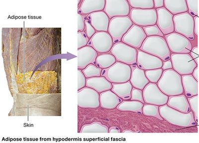
Adipose Tissue
Adipose tissue consists of adipocytes (fat-storing cells), providing insulation, protection, and energy storage.
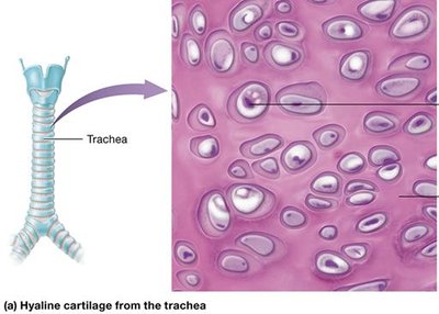
Cartilage
Cartilage is an avascular, tough, and flexible tissue found in joints, ears, nose, and respiratory passages. It contains chondroblasts and chondrocytes in lacunae.
Hyaline Cartilage: Most abundant, covers bone ends at joints.
Fibrocartilage: Found in intervertebral discs, strong and elastic.
Elastic Cartilage: Found in ear and larynx, handles vibration.
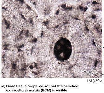
Bone (Osseous) Tissue
Bone tissue contains osteoblasts (build bone), osteocytes (maintain bone), and osteoclasts (resorb bone). The ECM is calcified for strength.
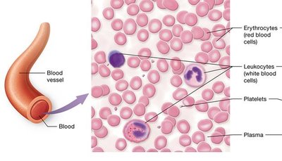
Blood
Blood is a fluid connective tissue with plasma as its ECM. It contains erythrocytes (oxygen transport), leukocytes (immunity), and platelets (clotting).