 Back
BackMini-Textbook Study Guide: The Integumentary System (Ch. 5)
Study Guide - Smart Notes
 Tailored notes based on your materials, expanded with key definitions, examples, and context.
The Integumentary System
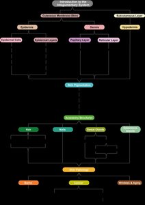
Introduction to the Integumentary System
The integumentary system is an organ system that includes the skin, hair, nails, glands, and sensory receptors. It is composed of several parts: the cutaneous membrane (skin), which consists of the epidermis and dermis, and the hypodermis (subcutaneous layer) beneath the skin. The system serves multiple essential functions for the body.
Protection: Acts as a barrier against mechanical stresses, chemicals, UV light, and microbes.
Homeostasis: Maintains internal stability, including temperature regulation.
Sensation: Contains nervous tissue for sensory perception.
Communication: Allows for expressive communication and emotions.
Vitamin D Synthesis: Skin synthesizes vitamin D when exposed to sunlight.
Excretion: Removes waste products via sweating.
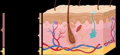
Structure of the Cutaneous Membrane (Skin)
Epidermis: The outermost layer, made of epithelial tissue.
Dermis: The deeper layer, composed of connective tissue.
Accessory Structures: Includes hair, nails, sweat glands, and sebaceous glands.
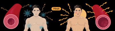
Thermoregulation in the Integumentary System
Mechanisms of Thermoregulation
The integumentary system is crucial for maintaining homeostasis, especially body temperature. Thermoregulation is achieved through two main methods:
Vasoconstriction & Vasodilation: Blood vessels in the dermis change diameter to regulate heat loss.
Sweating: Sweat glands secrete fluid that cools the body as it evaporates.
Vasoconstriction & Vasodilation
Vasoconstriction: Blood vessels constrict (decrease in diameter) when the body is cold, reducing blood flow to the skin and retaining heat.
Vasodilation: Blood vessels dilate (increase in diameter) when the body is hot, increasing blood flow to the skin and facilitating heat loss.
Sweating
Sweat: Water-based solution secreted by sweat glands when the body overheats.
Evaporation: Sweat evaporates from the skin surface, cooling the body.
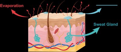
Example: After exercising on a hot day, vasodilation and active sweat glands cause a flushed appearance and cooling.
The Epidermis: Cells and Layers
Cell Types in the Epidermis
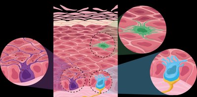
The epidermis is the outer layer of skin, composed of stratified squamous epithelial tissue. It contains four main cell types:
Keratinocytes: Most abundant; produce keratin, a tough, water-resistant protein.
Melanocytes: Produce melanin, a pigment that protects against UV damage.
Dendritic Cells (Langerhans cells): Immune cells that help prevent infection.
Tactile Epithelial Cells (Merkel cells): Specialized for touch sensation.

Keratinocytes
Connected by tight junctions (barrier) and desmosomes (mechanical strength).
Keratin provides mechanical and tensile strength, and is also found in hair and nails.
Example: Keratinocytes closer to the skin surface contain more keratin, making the skin tougher and more resistant.
Other Epidermal Cells
Melanocytes: Protect against UV radiation.
Dendritic Cells: Initiate immune responses.
Tactile Epithelial Cells: Detect touch.
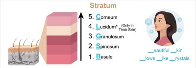
Layers of the Epidermis
The epidermis is composed of five distinct layers (strata), each with unique characteristics. The order from deep to superficial is:
Stratum Basale: Deepest layer; single row of proliferating cells.
Stratum Spinosum: Several rows of keratinocytes; thickest in thin skin.
Stratum Granulosum: Keratinocytes begin to harden and die; keratinization occurs.
Stratum Lucidum: Only in thick skin (palms, soles); clear, dead cells.
Stratum Corneum: Outermost layer; dead, keratin-filled cells.

Thin vs. Thick Skin
Thin Skin: Covers most of the body; lacks stratum lucidum; contains hair follicles and oil glands.
Thick Skin: Found on palms and soles; contains stratum lucidum; lacks hair follicles and oil glands; more sweat glands.

Keratinocyte Development
Keratinocytes originate in the stratum basale and are pushed upward through the layers, changing and dying as they move toward the surface.
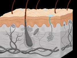
The Dermis
Structure and Layers of the Dermis
The dermis is the second layer of skin, located beneath the epidermis. It consists of two layers:
Papillary Layer: Superficial; made of loose connective tissue; contains dermal papillae, blood vessels, lymphatic vessels, and touch receptors (Meissner corpuscles).
Reticular Layer: Deep; made of dense irregular connective tissue; contains sweat and oil glands, hair roots, and pressure receptors (Pacinian corpuscles). Collagen and elastic fibers provide strength and flexibility.
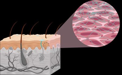
Dermal Papillae and Epidermal Ridges
Dermal Papillae: Projections that indent the epidermis, increasing surface area for attachment.
Epidermal Ridges: Enhance grip and produce fingerprints.

Reticular Layer Features
Collagen and Elastic Fibers: Provide strength and flexibility.
Cleavage Lines: Parallel arrangements of collagen fibers; incisions made parallel heal faster.
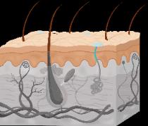
The Hypodermis (Subcutaneous Layer)
Structure and Functions of the Hypodermis
The hypodermis lies beneath the dermis and is not technically part of the skin. It is composed mostly of adipose tissue and areolar connective tissue. The hypodermis anchors the skin to underlying tissues, acts as a shock absorber, and insulates the body to reduce heat loss.
Anchoring: Attaches skin to muscles and bones.
Shock Absorption: Protects internal organs from mechanical injury.
Insulation: Helps maintain body temperature.
Fat Storage: Stores energy reserves.
Summary Table: Layers of the Skin
Layer | Main Features | Cell Types | Functions |
|---|---|---|---|
Epidermis | Stratified squamous epithelium; 5 strata | Keratinocytes, melanocytes, dendritic cells, tactile cells | Protection, water resistance, UV protection, sensation |
Dermis | Papillary (loose CT), Reticular (dense irregular CT) | Fibroblasts, immune cells, nerve endings | Strength, flexibility, sensation, blood supply |
Hypodermis | Adipose and areolar tissue | Adipocytes, fibroblasts | Anchoring, insulation, shock absorption, fat storage |
Key Equations and Concepts
Thermoregulation: Negative feedback loop maintains body temperature.
Keratinization: Process by which keratinocytes fill with keratin and die as they move toward the skin surface.
Additional info: The notes expand on brief points to provide full academic context, including definitions, examples, and comparisons. All images included are directly relevant to the adjacent content and reinforce understanding of skin structure and function.
