 Back
BackMini-Textbook Study Notes: Nervous Tissue and Nervous System
Study Guide - Smart Notes
 Tailored notes based on your materials, expanded with key definitions, examples, and context.
Nervous System: Histology and Organization
Overview of the Nervous System
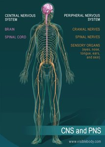
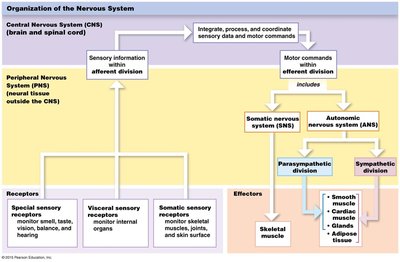
The nervous system is divided into two main anatomical and functional divisions: the Central Nervous System (CNS) and the Peripheral Nervous System (PNS). Each division plays a distinct role in communication, control, and integration of bodily functions.
Central Nervous System (CNS): Composed of the brain and spinal cord; responsible for processing and integrating sensory information and coordinating motor commands.
Peripheral Nervous System (PNS): Includes cranial nerves, spinal nerves, peripheral nerves, and sensory organs; gathers information from the environment and relays it to the CNS.
Nervous System Functions
The nervous system enables rapid communication and control throughout the body via electrical and chemical signals.
Communication: Electrical signals connect tissues to the CNS, allowing the body to sense and respond to external and internal stimuli.
Control: Adjusts activity of organs and tissues, producing swift but brief responses.
Conscious and Unconscious Awareness: Some sensory information reaches conscious awareness, while other signals (e.g., blood oxygen levels) are processed unconsciously.
Cellular Organization of Neural Tissue
Neurons and Neuroglia
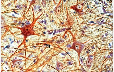
Neural tissue consists of two primary cell types:
Neurons: The basic functional unit responsible for transfer and processing of information. Neurons transmit signals via electrical impulses and chemical neurotransmitters. They consist of a cell body (soma), dendrites, and an axon.
Neuroglia (Glial Cells): Supporting cells that protect, nourish, and insulate neurons. Neuroglia outnumber neurons by approximately 10:1.
Structure of a Neuron
Neurons are specialized for communication and consist of several key components:
Dendrites: Receive information from other cells or the environment.
Cell Body (Soma): Contains the nucleus and organelles; site of metabolic activity.
Axon: Conducts nerve impulses away from the cell body toward synaptic terminals.
Terminal Endings: Release neurotransmitters to affect other neurons or effector organs.
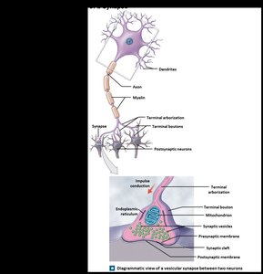
Synapses and Neuronal Communication
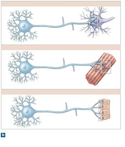
Synapses are specialized junctions where neurons communicate with other neurons, muscle fibers, or gland cells.
Presynaptic Neuron: Sends the signal.
Postsynaptic Neuron: Receives the signal.
Neurotransmitters: Chemical messengers released at the synapse to propagate the signal.
Classification of Neurons
Structural Classification
Neurons are classified based on their structure:
Anaxonic Neurons: Multiple processes; axons cannot be distinguished from dendrites. Found in the brain.
Bipolar Neurons: Two processes separated by the cell body. Associated with special senses (e.g., vision).
Pseudounipolar Neurons: Single elongated process with the cell body to one side. Most sensory neurons in the PNS.
Multipolar Neurons: Single axon and multiple dendrites. Most common motor neurons in the PNS.
Functional Classification
Neurons are also classified by the type of signals they convey:
Sensory Neurons: Transmit information from the PNS to the CNS. Includes somatic (conscious) and visceral (unconscious) sensory neurons.
Motor Neurons: Transmit information from the CNS to the periphery. Includes somatic (voluntary) and visceral (autonomic) motor neurons.
Interneurons: Located between sensory and motor neurons; analyze input and coordinate output. Can be excitatory or inhibitory.
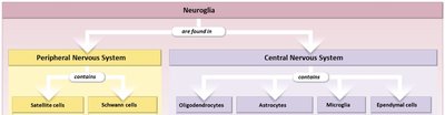
Neuroglia: Supporting Cells of Neural Tissue
Neuroglia in the CNS and PNS
Neuroglia are essential for the maintenance and function of neural tissue.
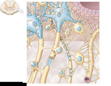
CNS Neuroglia: Astrocytes, oligodendrocytes, microglia, and ependymal cells.
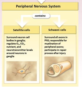
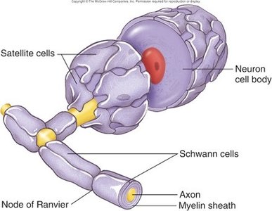
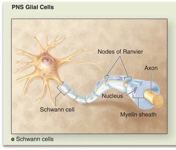
PNS Neuroglia: Satellite cells and Schwann cells.
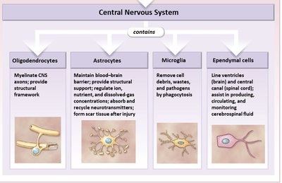
Neuroglia of the CNS
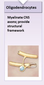
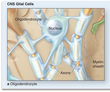
Oligodendrocytes: Myelinate CNS axons and provide structural framework.
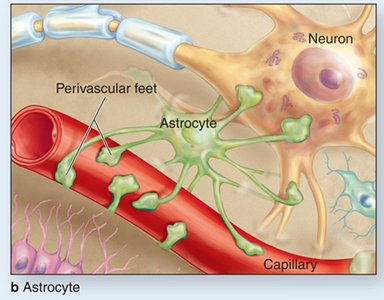
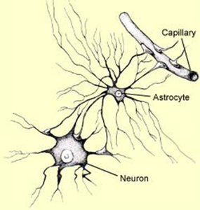
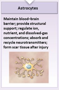
Astrocytes: Maintain the blood-brain barrier, provide structural support, regulate ion and nutrient concentrations, absorb/recycle neurotransmitters, and form scar tissue after injury.
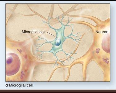
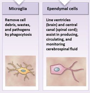
Microglia: Remove cell debris, wastes, and pathogens by phagocytosis.
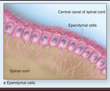
Ependymal Cells: Line ventricles and central canal; assist in producing, circulating, and monitoring cerebrospinal fluid (CSF).
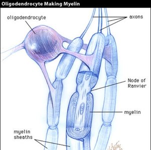
Oligodendrocytes and Myelination
Oligodendrocytes form myelin sheaths around axons in the CNS, which insulate the axon and speed up electrical signal transmission.
Myelin: Lipid-rich membrane that increases conduction velocity.
White Matter: Regions of the CNS rich in myelinated axons.
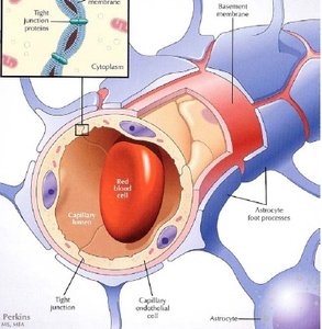
Astrocytes and the Blood-Brain Barrier
Astrocytes have specialized foot processes (perivascular feet) that cover capillaries in the CNS, forming the blood-brain barrier (BBB).
Blood-Brain Barrier: Protects nervous tissue by regulating the passage of substances from blood to brain.
Structural Support: Astrocytes hold neurons and capillaries together.
Regulation: Ensures only select molecules (O2, glucose, amino acids) enter the brain.

Microglia and Ependymal Cells
Microglia: Act as immune cells in the CNS, removing pathogens and debris.
Ependymal Cells: Line fluid-filled spaces (ventricles and central canal), produce and circulate cerebrospinal fluid (CSF).
Neuroglia of the PNS
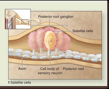
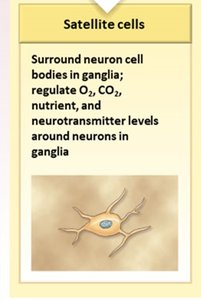
Satellite Cells: Surround neuron cell bodies in ganglia; regulate O2, CO2, nutrients, and neurotransmitter levels.
Schwann Cells: Surround all axons in the PNS; responsible for myelination and participate in repair after injury.
Summary Table: Neuroglia in CNS and PNS
Location | Cell Type | Main Function |
|---|---|---|
CNS | Astrocytes | Blood-brain barrier, structural support, regulation |
CNS | Oligodendrocytes | Myelination, structural framework |
CNS | Microglia | Immune defense, phagocytosis |
CNS | Ependymal cells | CSF production and circulation |
PNS | Satellite cells | Support neuron cell bodies, regulate environment |
PNS | Schwann cells | Myelination, repair |
Key Equations and Concepts
Action Potential
The transmission of electrical signals in neurons is governed by the action potential, described by the change in membrane potential:
Resting Membrane Potential:
Depolarization:
Propagation:
Myelination and Conduction Velocity
Myelinated axons conduct impulses faster due to saltatory conduction:
Conduction Velocity: (where is axon diameter)
Saltatory Conduction:
Conclusion
The nervous system is a highly organized network of neurons and neuroglia, each with specialized functions. Understanding the histology and cellular organization is fundamental to comprehending how the nervous system processes information and maintains homeostasis. Example: When you touch a hot object, sensory neurons transmit the signal to the CNS, interneurons process the information, and motor neurons initiate a rapid withdrawal response. Additional info: Academic context was added to clarify structural and functional classifications, synaptic mechanisms, and neuroglial functions.
