 Back
Mini-Textbook Study Notes: The Cardiovascular System – Heart Structure, Function, and Physiology
Study Guide - Smart Notes
 Tailored notes based on your materials, expanded with key definitions, examples, and context.
The Pericardium and Pericardial Cavity
Structure and Function of the Pericardium
The pericardium is a double-walled sac that surrounds the heart, providing protection, anchorage, and a frictionless environment for cardiac movement. It consists of two main layers: the fibrous pericardium and the serous pericardium.
Fibrous Pericardium: Tough, outer layer that prevents overstretching and anchors the heart to the diaphragm.
Serous Pericardium: Composed of two layers—parietal (lines the inner surface of the pericardial sac) and visceral (epicardium, covers the heart's surface).
Pericardial Cavity: Space between the parietal and visceral layers, filled with pericardial fluid to reduce friction during heartbeats.
Example: The pericardial cavity acts like a lubricated envelope, allowing the heart to beat smoothly within the thoracic cavity.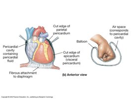
The Heart Wall
Layers of the Heart Wall
The heart wall is composed of three distinct layers, each with specialized functions.
Epicardium: The outermost layer, also known as the visceral pericardium.
Myocardium: The thick, middle layer made of cardiac muscle responsible for contraction.
Endocardium: The innermost layer, consisting of simple squamous epithelium, which lines the heart chambers and touches the blood.
Example: The myocardium is the functional muscle layer, enabling the heart to pump blood.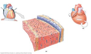
Cardiac Muscle Tissue
Structure and Special Features
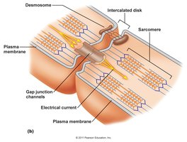
Cardiac muscle is unique among muscle tissues due to its involuntary, striated, and branched fibers. Cells are joined end-to-end by intercalated disks, which contain desmosomes and gap junctions.
Desmosomes: Physically join cells, providing structural integrity.
Gap Junctions: Form channels for ion flow, allowing rapid spread of action potentials and synchronized contraction.
Intercalated Disks: Specialized junctions that facilitate both mechanical and electrical connectivity.
Example: The heart contracts as a unit due to the rapid transmission of electrical impulses through gap junctions.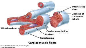
Gross Anatomy of the Heart
External and Internal Structures
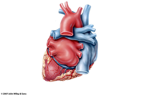
The heart is divided into four chambers: right and left atria (collecting chambers) and right and left ventricles (pumping chambers). The muscular septum separates the right and left sides.
Atria: Receive venous blood.
Ventricles: Eject arterial blood.
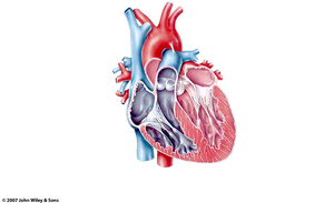
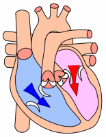
Valves: Atrioventricular (AV) valves separate atria and ventricles; semilunar valves separate ventricles from major arteries.
Example: The left ventricle has a thicker myocardium than the right, reflecting its role in pumping blood throughout the systemic circulation.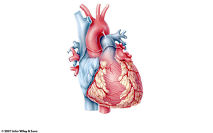
Heart Valves
Types and Functions
The heart contains four valves that ensure unidirectional blood flow:
Atrioventricular Valves: Tricuspid (right) and bicuspid/mitral (left) valves.
Semilunar Valves: Pulmonary (right) and aortic (left) valves.
Chordae Tendineae: Anchor AV valve cusps to papillary muscles, preventing inversion during contraction.
Example: AV valves open during relaxation, allowing blood to flow into ventricles; they close during contraction to prevent backflow.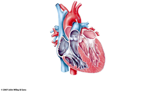
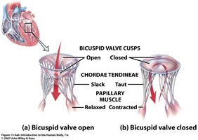
Valve Dynamics During Cardiac Cycle
Valves open and close in response to pressure changes between chambers and blood flow.
AV valves open when atrial pressure exceeds ventricular pressure.
Semilunar valves open when ventricular pressure exceeds arterial pressure.
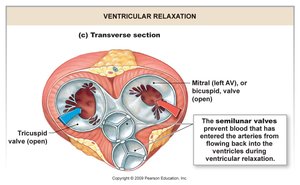
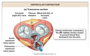
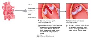
Path of Blood Flow Through the Heart
Sequential Blood Flow
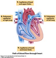
Blood flows through the heart in a specific sequence:
Right atrium → right ventricle → pulmonary artery → lungs
Lungs → pulmonary vein → left atrium → left ventricle → aortic valve
Aortic valve → aortic arch & coronary arteries → systemic circulation
Veins → superior/inferior vena cava → right atrium
Example: Oxygen-poor blood enters the right atrium, is pumped to the lungs, returns oxygen-rich to the left atrium, and is then pumped to the body.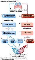
The Cardiac Cycle
Phases of the Cardiac Cycle
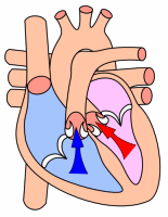
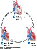
The cardiac cycle consists of three main phases:
Relaxation Period: All chambers are in diastole; ventricles fill with blood.
Atrial Systole: Atria contract, pushing the last 25% of blood into ventricles.
Ventricular Systole: Ventricles contract, ejecting blood into arteries.
Example: The cycle ensures continuous blood flow and efficient filling and emptying of heart chambers.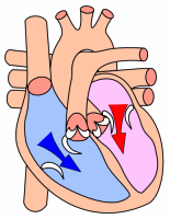
Coronary Circulation
Blood Supply to the Heart
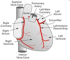
Coronary arteries originate at the base of the ascending aorta and supply blood to the myocardium. Venous blood from the myocardium returns via coronary veins.
Right and Left Coronary Arteries: Nourish heart tissue.
Coronary Veins: Drain deoxygenated blood from the myocardium.
Pacemaking and Conduction System
Electrical Conduction Pathway
The heart's rhythm is controlled by specialized pacemaker cells and a conduction system:
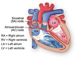
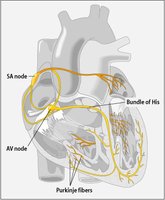
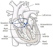
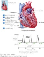
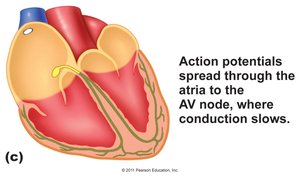
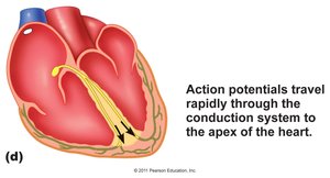
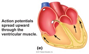
Sinoatrial (SA) Node: Initiates action potentials; natural pacemaker.
Atrioventricular (AV) Node: Delays impulse, allowing atrial contraction.
Bundle of His, Bundle Branches, Purkinje Fibers: Conduct impulses to ventricles, ensuring coordinated contraction.
Example: The SA node's intrinsic rate is ~100 beats/min, regulated by the autonomic nervous system.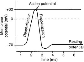
Electrophysiology of Cardiac Muscle
Action Potential Phases
Cardiac muscle action potentials have three phases:
Depolarization: Rapid Na+ influx via voltage-gated channels.
Plateau: Ca2+ influx (L-type channels) balances K+ outflow, maintaining depolarization.
Repolarization: Ca2+ channels close, K+ outflow restores resting membrane potential.
Equation:
Cardiac Output and Regulation
Determinants of Cardiac Output
Cardiac output is the volume of blood ejected per minute from the left ventricle. It is determined by heart rate and stroke volume.
Heart Rate (HR): Number of beats per minute, regulated by the SA node and autonomic nervous system.
Stroke Volume (SV): Volume of blood ejected per beat, influenced by preload, afterload, and contractility.
Equation:
Stroke Volume Influences
Preload: Degree of ventricular stretch during diastole.
Afterload: Resistance ventricles must overcome to eject blood.
Contractility: Force of ventricular contraction, dependent on Ca2+ and ATP availability.
Summary and Review
Key Concepts
The heart is a muscular organ with specialized structures for efficient blood flow.
Cardiac muscle features intercalated disks, desmosomes, and gap junctions for synchronized contraction.
Valves ensure unidirectional flow, opening and closing in response to pressure changes.
The cardiac cycle consists of relaxation, atrial systole, and ventricular systole.
Cardiac output is regulated by heart rate and stroke volume, influenced by neural and hormonal factors.
