 Back
Muscle Anatomy and Function: Study Notes for ANP College Students
Study Guide - Smart Notes
 Tailored notes based on your materials, expanded with key definitions, examples, and context.
Muscle Anatomy and Function
Basic Principles of Muscle Action
Muscles are responsible for producing movement by contracting and pulling on bones. Each muscle must cross a joint to move that joint, and has two points of attachment: the origin (stationary, usually proximal) and the insertion (mobile, usually distal).
Muscles only pull; they never push.
Movement occurs when a muscle contracts and pulls the insertion toward the origin.
Muscle Roles: Agonist, Antagonist, Synergist, Fixator
Muscles work together in groups to produce smooth and coordinated movements. The main roles are:
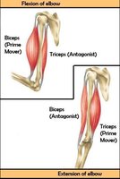
Agonist (Prime Mover): The muscle that produces the most force during a specific movement (e.g., biceps brachii during elbow flexion).
Antagonist: The muscle that opposes the prime mover (e.g., triceps brachii during elbow flexion).
Synergist: A muscle that assists the prime mover (e.g., deltoid in shoulder flexion).
Fixator: A muscle that stabilizes the origin of the prime mover (e.g., muscles stabilizing the scapula during arm movements).
Muscle Naming Conventions
Criteria for Naming Muscles
Muscles are named based on several characteristics:
Location: e.g., Biceps brachii (upper arm)
Number of Origins: e.g., "biceps" means two origins or heads
Origin and Insertion: e.g., Sternocleidomastoid (originates at sternum and clavicle, inserts on mastoid process)
Size: e.g., Gluteus maximus (largest gluteal muscle)
Shape: e.g., Deltoid (triangular shape)
Location of Flexors and Extensors
Flexors: Usually located on the ventral (anterior) side, except at the knee.
Extensors: Usually located on the dorsal (posterior) side, except at the knee.
Facial Muscles
Unique Features of Facial Muscles
Facial muscles typically insert into the skin rather than bone, allowing for a wide range of facial expressions. They are innervated by the Facial Nerve (Cranial Nerve VII).
Orbicularis oculi: Closes the eyes.
Orbicularis oris: Closes and puckers the mouth.
Zygomaticus major: Inserts into the corner of the mouth; responsible for smiling.
Buccinator: Compresses cheeks, holds food between teeth, and is used in whistling.
Mentalis: Pulls down the lower lip.
Facial Nerve Damage
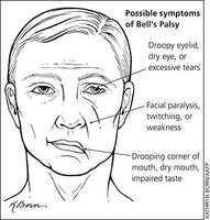
Damage to the facial nerve can result in flaccid paralysis of facial muscles, causing the face to droop on one side. Bell's Palsy is a common, usually reversible, condition involving facial nerve dysfunction.
Muscles of Mastication
Masseter
The masseter is a major muscle involved in chewing (mastication). It does not insert into the skin, but rather into the mandible, and is innervated by the Trigeminal Nerve (Cranial Nerve V).
Neck Muscles
Sternocleidomastoid and Scalenes
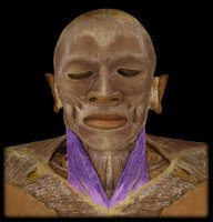
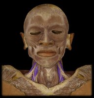
The sternocleidomastoid muscle flexes the neck and rotates the head to the opposite side. The scalenes are deep neck muscles that assist in breathing by elevating the rib cage.
Muscles of the Shoulder and Upper Back
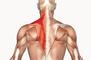
Trapezius
The trapezius muscle is a large, superficial muscle of the upper back and neck. It is involved in moving, rotating, and stabilizing the scapula and extending the neck.
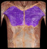
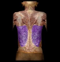
Pectoralis Major and Latissimus Dorsi
The pectoralis major is a large chest muscle responsible for flexion, adduction, and medial rotation of the humerus. The latissimus dorsi is a broad back muscle responsible for extension, adduction, and medial rotation of the shoulder.
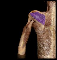
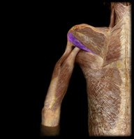
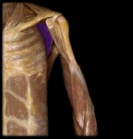
Rotator Cuff Muscles (SITS)
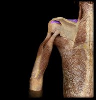
The rotator cuff is a group of four muscles that stabilize the shoulder joint and allow for circumduction and rotation:
Supraspinatus: Shoulder abduction
Infraspinatus: Lateral rotation of the shoulder
Teres minor: Lateral rotation and adduction
Subscapularis: Medial rotation
Scapular Stabilizers
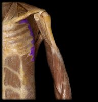
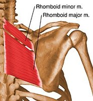
The rhomboid muscles stabilize the scapula, while the serratus anterior pulls the scapula forward and downward.
Deltoid
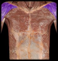
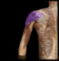
The deltoid is the prime mover for shoulder abduction. It has anterior (flexion), posterior (extension), and superior (abduction) fibers.
Muscles of the Upper Arm
Biceps Brachii
The biceps brachii has two heads (long and short) and crosses both the shoulder and elbow joints. It is responsible for flexion of the elbow and supination of the forearm.
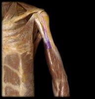
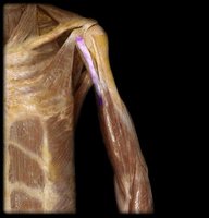
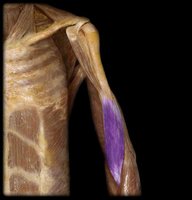
Brachialis
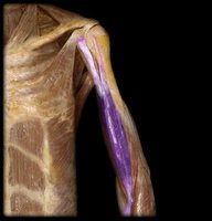
The brachialis lies deep to the biceps brachii and is a strong flexor of the elbow.
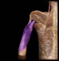
Triceps Brachii
The triceps brachii is the main extensor of the elbow joint.
Abdominal Muscles
Major Abdominal Muscles and Functions
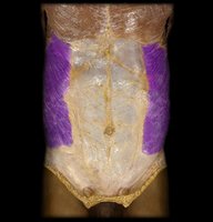
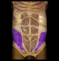
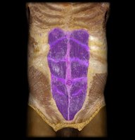
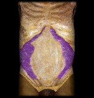
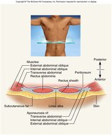
The abdominal wall is composed of several layers of muscle that provide trunk flexion, rotation, and compression of the abdomen:
External oblique: Compresses abdomen, rotates trunk
Internal oblique: Compresses abdomen, rotates trunk
Rectus abdominis: Flexes the vertebral column
Transverse abdominis: Compresses abdomen
Back Muscles
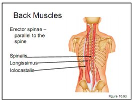
Erector Spinae
The erector spinae group (spinalis, longissimus, iliocostalis) runs parallel to the spine and is responsible for extending and laterally flexing the vertebral column.
Muscles of Respiration
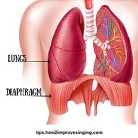
Diaphragm
The diaphragm is the primary muscle of breathing, contracting to increase thoracic volume and draw air into the lungs.
Hip Muscles
Flexors and Extensors
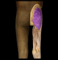
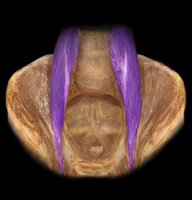
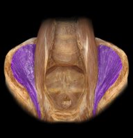
The iliopsoas (iliacus + psoas major) is the prime mover for hip flexion. The gluteus maximus is the prime mover for hip extension.
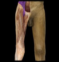
Hip Flexion Synergists
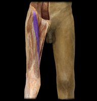
Sartorius: Spans two joints, assists in hip flexion and knee flexion.
Rectus femoris: Part of quadriceps femoris, also spans two joints, assists in hip flexion and knee extension.
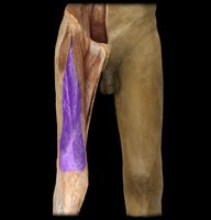
Quadriceps Femoris
The quadriceps femoris group consists of four muscles (rectus femoris, vastus lateralis, vastus medialis, vastus intermedius) and is the main extensor of the knee.
Hip Abductors and Adductors
Abductors (lateral thigh): Gluteus medius (prime mover), tensor fascia latae
Adductors (medial thigh): Adductor magnus, adductor longus, pectineus
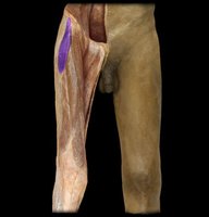
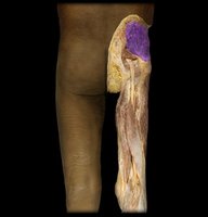
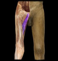
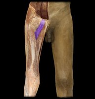
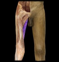
Iliotibial Tract (IT Band)
The iliotibial tract is a thick band of fascia running along the lateral thigh, providing stability to the knee and hip.
Hamstrings
Hamstring Muscles
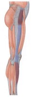
The hamstrings are located on the dorsal side of the thigh, span two joints, and are responsible for hip extension and knee flexion. The group includes:
Semitendinosus
Biceps femoris (lateral)
Semimembranosus
Antagonistic Muscle Groups
The hamstrings and quadriceps femoris are antagonistic muscle groups: hamstrings flex the knee and extend the hip, while quadriceps extend the knee and flex the hip.
Lower Leg Muscles
Major Muscles and Functions
Gastrocnemius: Flexes the knee and plantar flexes the ankle (via Achilles tendon).
Soleus: Plantar flexes the ankle (only).
Tibialis anterior: Dorsiflexes the ankle (anterior compartment).
Summary Table: Major Muscle Groups and Actions
Muscle Group | Primary Action | Location |
|---|---|---|
Biceps brachii | Elbow flexion, forearm supination | Anterior upper arm |
Triceps brachii | Elbow extension | Posterior upper arm |
Deltoid | Shoulder abduction | Shoulder |
Pectoralis major | Shoulder flexion, adduction, medial rotation | Chest |
Latissimus dorsi | Shoulder extension, adduction, medial rotation | Back |
Quadriceps femoris | Knee extension, hip flexion | Anterior thigh |
Hamstrings | Hip extension, knee flexion | Posterior thigh |
Gastrocnemius/Soleus | Plantar flexion of ankle | Posterior lower leg |
Tibialis anterior | Dorsiflexion of ankle | Anterior lower leg |
