 Back
BackMuscle and Epithelial Tissue Histology: Identification and Structure
Study Guide - Smart Notes
 Tailored notes based on your materials, expanded with key definitions, examples, and context.
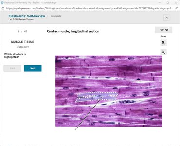
Q1. Which structure is highlighted in the cardiac muscle, longitudinal section?
Background
Topic: Cardiac Muscle Histology
This question tests your ability to identify key structures in cardiac muscle tissue under the microscope, specifically in a longitudinal section. Cardiac muscle is unique in its appearance and contains specialized junctions between cells.
Key Terms and Concepts:
Cardiac muscle fibers: Striated, branched cells found only in the heart.
Intercalated discs: Specialized connections between cardiac muscle cells that allow for synchronized contraction.
Step-by-Step Guidance
Observe the highlighted region in the image. Note the dark, distinct lines running perpendicular to the muscle fibers.
Recall that cardiac muscle cells are connected end-to-end by unique structures that appear as darker lines under the microscope.
Consider the function of these structures: they help transmit electrical impulses and maintain strong cell-to-cell adhesion during contraction.
Think about the name of these specialized junctions that are characteristic of cardiac muscle tissue.
Try solving on your own before revealing the answer!
Final Answer: Intercalated disc
The highlighted structure is an intercalated disc, which is a specialized junction between cardiac muscle cells. These discs are essential for the coordinated contraction of the heart.
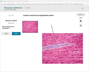
Q2. Which structure is highlighted in the cardiac muscle tissue, longitudinal section?
Background
Topic: Cardiac Muscle Histology
This question focuses on identifying the same or similar structures in a different section of cardiac muscle tissue. Recognizing these features is important for distinguishing cardiac muscle from other muscle types.
Key Terms and Concepts:
Cardiac muscle fibers
Intercalated discs
Step-by-Step Guidance
Examine the highlighted area for dark, transverse lines crossing the muscle fibers.
Recall the function and appearance of intercalated discs in cardiac muscle tissue.
Compare the structure to those seen in skeletal or smooth muscle to confirm your identification.
Try solving on your own before revealing the answer!
Final Answer: Intercalated disc
The highlighted structure is an intercalated disc, which is unique to cardiac muscle and is critical for cell-to-cell communication and contraction.
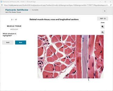
Q3. Which structure is highlighted in the skeletal muscle tissue; cross and longitudinal sections?
Background
Topic: Skeletal Muscle Histology
This question asks you to identify a structure in both cross and longitudinal sections of skeletal muscle. Understanding the orientation and appearance of muscle fibers is key in histology.
Key Terms and Concepts:
Skeletal muscle fiber: Long, cylindrical, multinucleated cells with striations.
Longitudinal section: Shows the length of the muscle fibers.
Cross section: Shows the muscle fibers in cross-cut, appearing as circles.
Step-by-Step Guidance
Look at the highlighted area and determine if it is showing the length or the cross-section of the muscle fiber.
Recall the appearance of skeletal muscle fibers in both orientations: striations in longitudinal, circular profiles in cross-section.
Identify the structure based on its orientation and appearance.
Try solving on your own before revealing the answer!
Final Answer: Skeletal muscle fiber, longitudinal section
The highlighted structure is a skeletal muscle fiber seen in longitudinal section, showing the characteristic striations of skeletal muscle.
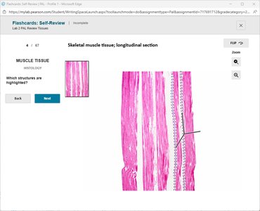
Q4. Which structure is highlighted in the skeletal muscle tissue; longitudinal section?
Background
Topic: Skeletal Muscle Histology
This question focuses on identifying the muscle fiber in a longitudinal section of skeletal muscle tissue. Recognizing the striated appearance is important for distinguishing skeletal muscle from other types.
Key Terms and Concepts:
Skeletal muscle fiber
Striations
Step-by-Step Guidance
Observe the highlighted region for long, parallel fibers with visible striations.
Recall that skeletal muscle fibers are multinucleated and show a striped pattern due to the arrangement of actin and myosin filaments.
Identify the structure based on these features.
Try solving on your own before revealing the answer!
Final Answer: Skeletal muscle fiber, longitudinal section
The highlighted structure is a skeletal muscle fiber in longitudinal section, showing the typical striations of skeletal muscle.
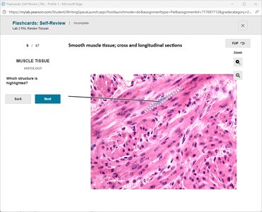
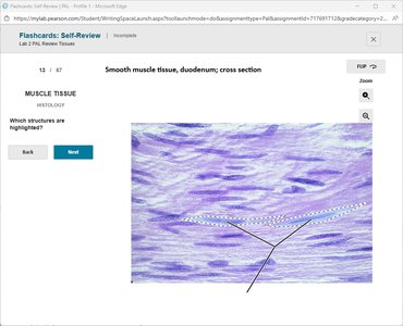
Q5. Which structure is highlighted in the smooth muscle tissue; cross and longitudinal sections?
Background
Topic: Smooth Muscle Histology
This question tests your ability to identify smooth muscle fibers in both cross and longitudinal sections. Smooth muscle is found in the walls of hollow organs and has a distinct appearance compared to skeletal and cardiac muscle.
Key Terms and Concepts:
Smooth muscle fiber: Spindle-shaped, non-striated cells with a single central nucleus.
Longitudinal section: Shows elongated fibers.
Cross section: Shows small, round to oval profiles.
Step-by-Step Guidance
Examine the highlighted area for elongated, non-striated fibers (longitudinal) or small, round profiles (cross-section).
Recall that smooth muscle lacks the striations seen in skeletal and cardiac muscle.
Identify the structure based on its shape and lack of striations.
Try solving on your own before revealing the answer!
Final Answer: Smooth muscle fibers, longitudinal section
The highlighted structure is a smooth muscle fiber in longitudinal section, characterized by its elongated shape and lack of striations.
Q6. Which epithelial type is highlighted in the trachea cross section?
Background
Topic: Epithelial Tissue Histology
This question asks you to identify the type of epithelial tissue lining the trachea. Recognizing epithelial types is essential for understanding tissue function and location.
Key Terms and Concepts:
Pseudostratified columnar epithelium: Appears to have multiple layers due to nuclei at different levels, but all cells touch the basement membrane.
Location: Commonly found lining the respiratory tract, including the trachea.
Step-by-Step Guidance
Observe the highlighted region for tall, column-shaped cells with nuclei at varying heights.
Recall that this epithelium often contains cilia and goblet cells for mucus production and movement.
Identify the tissue based on these features and its location in the trachea.
Try solving on your own before revealing the answer!
Final Answer: Pseudostratified columnar epithelium
The highlighted tissue is pseudostratified columnar epithelium, which lines the trachea and is specialized for secretion and movement of mucus.
