 Back
Muscle Contraction and Metabolism: Study Notes for ANP College Students
Study Guide - Smart Notes
 Tailored notes based on your materials, expanded with key definitions, examples, and context.
Whole Muscle Contraction
Motor Units and Neuromuscular Junctions
Muscle contraction is controlled by motor nerves, which contain axons of motor neurons. Each axon branches into terminals that form neuromuscular junctions (NMJs) with individual muscle fibers. The motor unit is the fundamental functional unit, consisting of a motor neuron and all the muscle fibers it innervates. Motor units can be small (few fibers, fine control) or large (hundreds of fibers, gross movements).
Motor Unit: A single motor neuron and all the muscle fibers it controls.
Neuromuscular Junction: The synapse between a motor neuron and a muscle fiber.
Fine vs. Gross Control: Small motor units allow precise movements; large units enable powerful contractions.
Example: Muscles controlling eye movements have small motor units; leg muscles have large motor units.
Muscle Metabolism
ATP and Its Role in Muscle Contraction
ATP is the primary energy source for muscle contraction. It is required for moving and detaching cross bridges, pumping calcium back into the sarcoplasmic reticulum (SR), and restoring sodium and potassium gradients after excitation-contraction coupling. Stored ATP is depleted within 4–6 seconds, so it must be regenerated rapidly.
ATP Functions: Cross-bridge cycling, calcium reuptake, ion gradient restoration.
Regeneration: ATP is regenerated by three main pathways.
Pathways for ATP Synthesis
Muscle fibers regenerate ATP through three mechanisms:
Direct Phosphorylation: Creatine phosphate (CP) donates a phosphate to ADP, forming ATP. This process is catalyzed by creatine kinase and provides energy for about 15 seconds.
Anaerobic Pathway: Glycolysis breaks down glucose (from glycogen or blood) to produce ATP and pyruvic acid. In the absence of oxygen, pyruvic acid is converted to lactic acid, which diffuses into the bloodstream and is eventually converted back to glucose in the liver.
Aerobic Pathway: Aerobic respiration occurs in mitochondria, using glucose, fatty acids, and amino acids to produce ATP, CO2, and H2O. This pathway is slower but produces most ATP during rest and prolonged exercise.
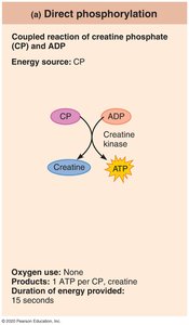
Summary Table: ATP Regeneration Pathways
Direct Phosphorylation: Fast, no oxygen required, limited duration.
Anaerobic Glycolysis: Moderate speed, no oxygen required, produces lactic acid.
Aerobic Respiration: Slow, requires oxygen, sustained energy production.
Energy Pathways During Exercise
Different types of exercise utilize different energy pathways:
Short-duration, high-intensity exercise: Uses stored ATP, then creatine phosphate, then anaerobic glycolysis.
Prolonged-duration exercise: Relies on aerobic respiration for sustained ATP production.
Example: Sprinting uses anaerobic pathways; marathon running uses aerobic pathways.
Muscle Fiber Types
Classification of Muscle Fibers
Muscle fibers are classified based on their speed of contraction and metabolic pathways used for ATP synthesis. The three main types are:
Slow Oxidative Fibers: Use aerobic pathways, fatigue-resistant, suited for endurance activities.
Fast Oxidative Fibers: Use aerobic and some anaerobic pathways, intermediate fatigue resistance, suited for sprinting and walking.
Fast Glycolytic Fibers: Use anaerobic glycolysis, fatigue quickly, suited for short-term intense movements.
Functional Characteristics and Activities
Slow Oxidative: Low-intensity, endurance (e.g., maintaining posture).
Fast Oxidative: Medium-intensity (e.g., sprinting, walking).
Fast Glycolytic: High-intensity, short duration (e.g., hitting a baseball).
Genetics: Determines individual distribution of fiber types.
Structural and Functional Characteristics Table
The following table summarizes the structural and functional characteristics of the three types of skeletal muscle fibers:
Characteristic | Slow Oxidative Fibers | Fast Oxidative Fibers | Fast Glycolytic Fibers |
|---|---|---|---|
Speed of Contraction | Slow | Fast | Fast |
Myosin ATPase Activity | Slow | Fast | Fast |
Primary ATP Pathway | Aerobic | Aerobic (some anaerobic) | Anaerobic glycolysis |
Myoglobin Content | High | High | Low |
Glycogen Stores | Low | Intermediate | High |
Recruitment Order | First | Second | Third |
Fatigue Resistance | High | Intermediate | Low |
Best Suited For | Endurance (posture, running) | Sprinting, walking | Short-term intense (hitting a baseball) |
Fiber Diameter | Small | Large | Intermediate |
Mitochondria | Many | Many | Few |
Capillaries | Many | Many | Few |
Color | Red | Red to pink | White |
Additional info: The table above is inferred from Table 9.2 and summarizes the main differences between muscle fiber types, including their metabolic and structural properties.
