 Back
BackMuscle Fiber Energy Sources and Tension Production
Study Guide - Smart Notes
 Tailored notes based on your materials, expanded with key definitions, examples, and context.
CH 10 PT 5 - Muscle Fiber Energy Sources
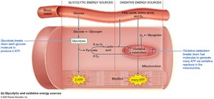
Glycolytic (Anaerobic) Catabolism
Glycolytic catabolism is a series of reactions that occur in the cytosol of muscle fibers, where a six-carbon glucose molecule is split into two three-carbon pyruvate molecules. This process produces a net gain of two ATP molecules per glucose and does not require oxygen directly.
Glucose Sources: Muscle fibers obtain glucose from the bloodstream and from glycogen stores within the muscle (and liver).
Duration: Glycolysis can provide adequate ATP for about 30–40 seconds of sustained muscle contraction.
Fate of Pyruvate: If oxygen is abundant, pyruvate enters the mitochondria for oxidative catabolism. If oxygen is scarce, pyruvate is converted to lactic acid, some of which is transported to the liver and converted back to glucose.
Oxidative (Aerobic) Catabolism
Oxidative catabolism occurs in the mitochondria and involves the removal of electrons from carbon-based compounds, using the energy released to synthesize ATP. This process requires oxygen and provides the majority of ATP for sustained muscle activity.
Fuel Sources: Muscle fibers use glucose first, then fatty acids and amino acids as glucose becomes less available.
Oxygen Supply: Oxygen is supplied from the bloodstream and from myoglobin, an oxygen-binding protein in muscle fibers.
Duration: Oxidative catabolism can provide ATP for hours, becoming the predominant source after about 1 minute of activity.
Muscle Tension at the Fiber Level
Muscle Twitch
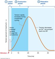
A muscle twitch is the response of a muscle fiber to a single action potential from a motor neuron, representing the smallest possible muscle contraction. Twitches are typically observed in laboratory settings with direct electrical stimulation.
Myogram: A recording of a muscle twitch, showing the changes in tension over time.
Phases of a Twitch:
Latent Period: 1–2 ms; action potential spreads through the sarcolemma, leading up to crossbridge cycling.
Contraction Period: Tension increases rapidly as crossbridge cycles occur.
Relaxation Period: Tension decreases as calcium ions are pumped back into the sarcoplasmic reticulum (SR).
Refractory Period: About 5 ms during which the muscle fiber cannot respond to another stimulus.
Tension Production and the Timing and Frequency of Stimulation
Wave Summation
Wave summation occurs when a muscle fiber is repeatedly stimulated by a motor neuron, resulting in twitches with progressively greater tension. This is due to incomplete removal of calcium ions from the cytosol before subsequent stimuli.
Unfused (Incomplete) Tetanus: Stimulation at about 50 times per second allows only partial relaxation between contractions, causing tension to pulsate.
Fused (Complete) Tetanus: Stimulation at more than 80–100 times per second prevents relaxation, resulting in a sustained contraction and maximal tension.
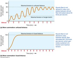
Unfused and fused tetanus generate more tension than a single twitch, with fused tetanus producing the highest tension. The bacterium Clostridium tetani can cause excessive muscle contraction (tetanic spasm) by releasing a toxin that overstimulates skeletal muscles.
Botulism and Botox
Clostridium botulinum and Botulinum Toxin
Clostridium botulinum produces the most lethal known biological toxin, which can block acetylcholine release at the neuromuscular junction, preventing muscle contraction and potentially causing death by respiratory failure. The toxin is most commonly encountered through improperly sterilized canned foods.
In medicine, small doses of botulinum toxin (Botox®) are used therapeutically to relieve muscle spasms, treat migraines, and for cosmetic purposes to reduce wrinkles by relaxing facial muscles.
The Length-Tension Relationship
Relationship Between Sarcomere Length and Tension
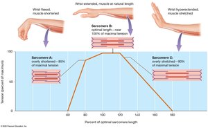
The amount of tension a muscle fiber can produce depends on the length of its sarcomeres prior to contraction. The optimal length allows for the greatest number of crossbridges to form, resulting in maximal tension.
Overly Shortened Muscle: Excessive overlap of filaments limits tension production.
Overly Stretched Muscle: Minimal overlap prevents myosin heads from effectively binding to actin, reducing tension.
Optimal Length: Ideal overlap allows for maximal crossbridge formation and tension generation.
Classes of Skeletal Muscle Fibers
Fast-Twitch and Slow-Twitch Fibers
Skeletal muscle fibers are classified based on their speed of contraction and predominant energy source.
Fast-Twitch Fibers: High myosin ATPase activity; rapid contractions; found in muscles responsible for quick movements (e.g., eye muscles).
Slow-Twitch Fibers: Low myosin ATPase activity; slower, sustained contractions; found in postural muscles.
Type I and Type II Fibers
Type I (Slow Oxidative) Fibers: Small to intermediate diameter; contract slowly with less force but resist fatigue; high myoglobin content ("red muscle"), many mitochondria, and rich blood supply.
Type II (Fast Glycolytic) Fibers: Larger diameter; contract rapidly but fatigue quickly; rely on glycolytic energy sources; low myoglobin content ("white muscle"), fewer mitochondria, and less extensive blood supply.
