 Back
BackMuscle Performance and Muscle Tissue Types in Human Anatomy & Physiology
Study Guide - Smart Notes
 Tailored notes based on your materials, expanded with key definitions, examples, and context.
Muscle Performance
Key Concepts in Muscle Performance
Muscle performance is a critical aspect of human anatomy and physiology, often evaluated in terms of force (power, strength) and endurance. Force refers to the maximum tension produced by a muscle or muscle group, while endurance is the duration over which a muscle can sustain activity.
Force (Power, Strength): The maximum tension a muscle can generate.
Endurance: The length of time a muscle can maintain activity.
Factors Affecting Performance: Number of muscle fibers, physical conditioning, and genetic predisposition.
Types of Skeletal Muscle Fibers
Skeletal muscle fibers are classified based on their contraction speed, metabolic properties, and resistance to fatigue. The three main types are slow fibers (Type I), fast fibers (Type II-B), and intermediate fibers (Type II-A).
Slow Fibers (Type I): Also known as slow twitch oxidative fibers, these are fatigue-resistant and suited for endurance activities.
Fast Fibers (Type II-B): Fast twitch glycolytic fibers, large in diameter, produce rapid, powerful contractions but fatigue quickly.
Intermediate Fibers (Type II-A): Fast twitch oxidative fibers, with properties between slow and fast fibers, more resistant to fatigue than fast fibers.

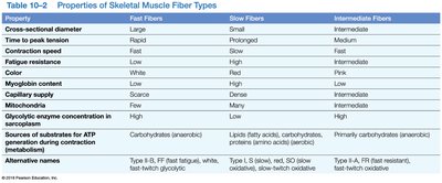
Properties of Skeletal Muscle Fiber Types
The following table summarizes the main properties of fast, slow, and intermediate muscle fibers:
Property | Fast Fibers | Slow Fibers | Intermediate Fibers |
|---|---|---|---|
Cross-sectional diameter | Large | Small | Intermediate |
Time to peak tension | Rapid | Prolonged | Medium |
Contraction speed | Fast | Slow | Fast |
Fatigue resistance | Low | High | Intermediate |
Color | White | Red | Pink |
Myoglobin content | Low | High | Intermediate |
Capillary supply | Scarce | Many | Intermediate |
Mitochondria | Few | Many | Intermediate |
Glycolysis enzyme concentration | High | Low | High |
Sources of substrates for ATP | Carbohydrates | Lipids, fatty acids, carbohydrates, proteins | Primarily carbohydrates |
Alternative names | Type II-B, fast fatigue, white, fast twitch glycolytic | Type I, slow, red, slow twitch oxidative | Type II-A, fast fatigue resistant, fast twitch oxidative |
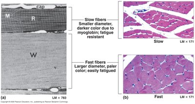
Microscopic Comparison of Muscle Fibers
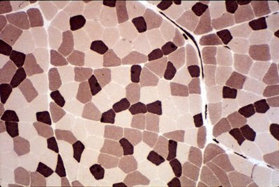
Microscopic images reveal differences in muscle fiber size and color, reflecting their myoglobin content and fatigue resistance.
Slow fibers: Smaller diameter, darker color due to high myoglobin, fatigue resistant.
Fast fibers: Larger diameter, paler color, easily fatigued.
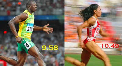
Muscle Performance and Endurance
Aerobic vs. Anaerobic Endurance
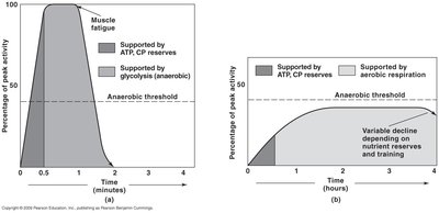
Muscle endurance is supported by different metabolic pathways depending on activity intensity and duration. Anaerobic endurance relies on glycolysis and ATP/CP reserves for brief, intense activity, while aerobic endurance depends on sustained oxygen supply and aerobic metabolism for prolonged activity.
Anaerobic endurance: Short, intense activity; increases muscle mass, ATP/CP, and glycogen reserves.
Aerobic endurance: Long-duration activity; increases blood supply, cardiovascular efficiency, and mitochondrial content.


Muscle Hypertrophy and Atrophy
Hypertrophy
Muscle hypertrophy is the increase in muscle cell diameter due to synthesis of more actin and myosin myofilaments, typically resulting from increased activity and training.
Hypertrophy: Increase in muscle cell diameter; existing cells get bigger.
Mechanism: Synthesis of more myofilaments (actin and myosin).
Atrophy
Muscle atrophy is the decrease in muscle cell diameter due to loss of myofilaments, often caused by disuse, immobility, or nerve damage. Long-term atrophy can be irreversible if muscle cells die.
Atrophy: Decrease in muscle cell diameter; loss of myofilaments.
Causes: Disuse, immobility, nerve damage.
Aging and the Muscular System
Effects of Aging
Aging leads to a decrease in muscle size, strength, and endurance, primarily due to a reduction in myofibrils, ATP, CP, glycogen, and myoglobin. Muscles become less elastic and more fibrous, with reduced repair capabilities.
Decrease in myofibrils: Reduced muscle size and strength.
Reduced endurance: Less ATP, CP, glycogen, and myoglobin.
Fibrosis: Increased fibrous connective tissue.
Reduced repair: Fewer satellite cells, more scar tissue formation.
Muscular System Disorders
Primary Disorders
Primary muscular disorders originate within the muscle itself and include trauma, infections, inherited disorders, and tumors.
Muscle trauma: Damage from excessive activity or injury.
Muscle infections: Inflammation due to pathogens.
Inherited disorders: Muscular dystrophies (e.g., Duchenne's, myotonic dystrophy).
Tumors: Sarcoma.
Secondary Disorders
Secondary disorders result from problems in other systems, such as nervous, metabolic, or cardiovascular systems, affecting muscle function.
Nervous system disorders: Affect coordination or control of muscle contraction.
Metabolic/nutritional disorders: Affect energy supply or electrolyte balance.
Cardiovascular disorders: Restrict blood flow to muscles.
Common Muscular Disorders
Muscle spasm (cramp): Strong, sudden, painful contraction.
Muscle spasticity: Excessive muscle tone.
Muscle flaccidity: Very low muscle tone.
Muscle atrophy: Deterioration due to disuse or nerve damage.
Myositis: Muscle inflammation (autoimmune types: polymyositis, dermatomyositis).
Strain: Tears in muscle tissue.
Sprain: Tears in ligaments, tendons, or joint capsules.
Paralysis: Loss of voluntary motor control (flaccid or spastic).
Muscle Tissue Types
Skeletal Muscle
Skeletal muscle is striated, voluntary, and attached to bones. It is responsible for body movement and posture.
Striated: Light/dark bands due to sarcomeres.
Voluntary control: Controlled by motor neurons.
Multinucleate: Formed by fusion of myoblasts.
Cardiac Muscle
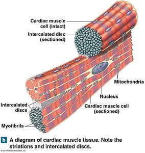
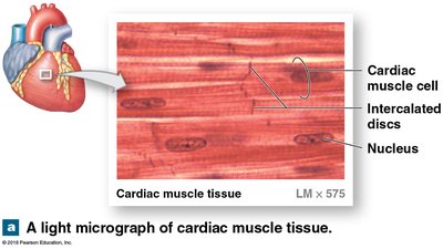
Cardiac muscle forms the walls of the heart, is striated and involuntary, and features intercalated discs for electrical and structural connectivity.
Striated: Actin and myosin arranged in sarcomeres.
Involuntary control: Controlled by pacemaker cells and autonomic nervous system.
Intercalated discs: Structural and electrical connections between cells.
Smooth Muscle
Smooth muscle is non-striated, involuntary, and found in the walls of hollow internal organs. It is responsible for movements such as blood vessel constriction, digestive tract motility, and uterine contractions.
Non-striated: Actin and myosin not organized in sarcomeres.
Involuntary control: Controlled by pacesetter cells, hormones, and autonomic nervous system.
Plasticity: Ability to adapt to new lengths and retain contractile function.
Comparison of Muscle Tissue Types
Size and Structure
Skeletal: Diameter ~100 µm, length up to 30 cm.
Cardiac: Diameter 10-20 µm, length 50-100 µm.
Smooth: Diameter 5-10 µm, length 30-200 µm.
Filament Organization
Skeletal & Cardiac: Striated, actin & myosin in sarcomeres.
Smooth: Non-striated, actin & myosin not in sarcomeres.
Control Mechanisms
Skeletal: Voluntary, motor neurons.
Cardiac: Involuntary, pacemaker cells, autonomic nervous system.
Smooth: Involuntary, pacesetter cells, hormones, autonomic nervous system.
Energy Source
Skeletal: Aerobic at moderate activity, anaerobic at peak.
Cardiac: Aerobic metabolism, high myoglobin and mitochondria.
Smooth: Aerobic at moderate activity, anaerobic at peak (rare).
Contraction Types
Skeletal: Tetanic contractions produce greatest tension.
Cardiac: Twitch contractions only.
Smooth: Tetanic contractions, summation of tension.
Key Equations
Muscle contraction and energy production involve several biochemical equations:
ATP Hydrolysis: $\mathrm{ATP} + \mathrm{H}_2\mathrm{O} \rightarrow \mathrm{ADP} + \mathrm{P}_i + \text{energy}$
Glycolysis (Anaerobic): $\mathrm{C}_6\mathrm{H}_{12}\mathrm{O}_6 \rightarrow 2\mathrm{C}_3\mathrm{H}_6\mathrm{O}_3 + 2\mathrm{ATP}$
Aerobic Respiration: $\mathrm{C}_6\mathrm{H}_{12}\mathrm{O}_6 + 6\mathrm{O}_2 \rightarrow 6\mathrm{CO}_2 + 6\mathrm{H}_2\mathrm{O} + 36\mathrm{ATP}$
Additional info: These equations represent the primary pathways for energy production in muscle cells, supporting both short-term and long-term muscle activity.
