 Back
BackMuscle Performance, Fiber Types, and Muscle Tissue Comparison
Study Guide - Smart Notes
 Tailored notes based on your materials, expanded with key definitions, examples, and context.
Muscle Performance
Key Concepts in Muscle Performance
Muscle performance is evaluated based on two primary factors: force (power, strength) and endurance. Force refers to the maximum tension a muscle or muscle group can produce, while endurance is the duration a muscle can sustain activity.
Force (Power, Strength): The peak tension generated by a muscle or group.
Endurance: The length of time a muscle can perform a specific activity.
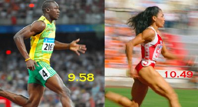
Determinants of Performance: The proportion of different muscle fiber types and the effects of physical conditioning or training.
Types of Skeletal Muscle Fibers
Classification and Characteristics
Skeletal muscle fibers are classified into three main types based on their contraction speed, metabolic properties, and resistance to fatigue:
Slow Fibers (Type I, Slow Twitch Oxidative): Adapted for endurance and continuous contraction. They are rich in mitochondria and myoglobin, use aerobic metabolism, and are fatigue-resistant.
Fast Fibers (Type II-B, Fast Twitch Glycolytic): Adapted for rapid, powerful contractions of short duration. They have large diameters, high glycogen reserves, and rely on anaerobic metabolism, making them prone to fatigue.
Intermediate Fibers (Type II-A, Fast Twitch Oxidative): Exhibit properties between slow and fast fibers. They are fast-contracting but have greater resistance to fatigue than fast fibers due to increased mitochondrial content and capillary supply.

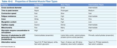
Comparison Table: Properties of Skeletal Muscle Fiber Types
The following table summarizes the main differences among fast, slow, and intermediate muscle fibers:
Property | Fast Fibers | Slow Fibers | Intermediate Fibers |
|---|---|---|---|
Cross-sectional diameter | Large | Small | Intermediate |
Time to peak tension | Rapid | Prolonged | Medium |
Contraction speed | Fast | Slow | Fast |
Fatigue resistance | Low | High | Intermediate |
Color | White | Red | Pink |
Myoglobin content | Low | High | Low |
Capillary supply | Scarce | Dense | More than fast, less than slow |
Mitochondria | Few | Many | More than fast, less than slow |
Glycolytic enzyme concentration | High | Low | High |
Sources of substrates for ATP | Carbohydrates (anaerobic) | Lipids, carbohydrates, proteins (aerobic) | Primarily carbohydrates (anaerobic) |
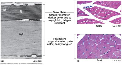
Microscopic and Histological Differences
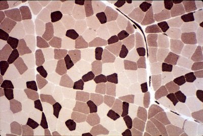
Slow fibers appear darker due to higher myoglobin content, while fast fibers are paler and larger in diameter. These differences are visible under the microscope and are linked to their functional properties.
Muscle Hypertrophy and Atrophy
Changes in Muscle Size
Muscle hypertrophy is the increase in muscle fiber diameter due to increased synthesis of actin and myosin myofilaments, typically from resistance training. Atrophy is the decrease in muscle fiber diameter due to disuse, immobilization, or loss of neural stimulation. The number of muscle fibers does not change in adults; only their size varies.
Hypertrophy: Increased muscle size from more myofilaments.
Atrophy: Decreased muscle size from loss of myofilaments; can become irreversible if muscle cells die.
Muscle Performance and Endurance
Aerobic vs. Anaerobic Endurance
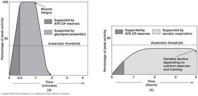
Endurance is supported by different metabolic pathways depending on the intensity and duration of activity:
Anaerobic Endurance: Short, intense activities rely on ATP and creatine phosphate (CP) reserves, then glycolysis (anaerobic metabolism).
Aerobic Endurance: Prolonged, moderate activities rely on aerobic metabolism, supported by oxygen delivery and mitochondrial function.
Physical Conditioning
Anaerobic Training: Increases muscle mass, ATP/CP and glycogen reserves, and tolerance to lactic acid (e.g., weightlifting, sprinting).
Aerobic Training: Increases blood supply, cardiovascular efficiency, and mitochondrial density (e.g., jogging, cycling).


Aging and the Muscular System
Effects of Aging
With age, muscles decrease in size, strength, and endurance due to loss of myofibrils, reduced ATP/CP/glycogen/myoglobin, and increased fibrous tissue (fibrosis). Recovery from injury is slower, and the number of satellite cells declines, limiting repair capacity.
Muscular System Disorders
Primary and Secondary Disorders
Primary Disorders: Directly affect muscle tissue (e.g., trauma, infections, inherited disorders like muscular dystrophy, tumors).
Secondary Disorders: Originate in other systems but impact muscle function (e.g., nervous, metabolic, cardiovascular disorders).
Common Disorders and Symptoms
Muscle Spasm (Cramp): Sudden, painful contraction.
Spasticity: Excessive muscle tone.
Flaccidity: Very low muscle tone.
Atrophy: Wasting due to disuse or nerve damage.
Myositis: Inflammation of muscle tissue (autoimmune or infectious).
Strain: Muscle tear; Sprain: Ligament/tendon/joint capsule tear.
Paralysis: Loss of voluntary control (flaccid or spastic).
Nervous System Disorders Affecting Muscle
Blockage of Acetylcholine (ACh) Release: Flaccid paralysis (e.g., botulism).
Interference with ACh Binding: Flaccid paralysis (e.g., autoimmune diseases).
Interference with ACh Esterase: Spastic paralysis (e.g., organophosphates, nerve gas).
Loss of Motor Neurons: Flaccid paralysis (e.g., polio).
Peripheral Nerve Damage: Flaccid paralysis.
Excessive Stimulation (e.g., tetanus): Spastic paralysis.
Inherited Muscle Disorders
Duchenne's Muscular Dystrophy: X-linked, early onset, more common in males.
Myotonic Dystrophy: Chromosome 19, onset after puberty, affects both sexes equally.
Cardiac and Smooth Muscle
Overview of Muscle Tissue Types
All muscle tissues are specialized for contraction but differ in structure, control, and function:
Skeletal Muscle: Striated, voluntary, attached to bones.
Cardiac Muscle: Striated, involuntary, forms heart walls.
Smooth Muscle: Non-striated, involuntary, found in walls of hollow organs.
Cardiac Muscle Structure
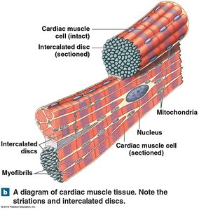
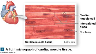
Cardiac muscle cells are connected by intercalated discs containing desmosomes and gap junctions, allowing electrical and mechanical coupling for synchronized contraction.
Smooth Muscle Structure and Function
Smooth muscle cells are small, spindle-shaped, and lack striations. They are found in the walls of blood vessels, digestive, respiratory, urinary, and reproductive organs. Contraction is controlled by autonomic nervous system, hormones, and pacemaker cells.
Plasticity: Ability to adapt to stretching and maintain contractile function.
Myofilament Organization: Thick and thin filaments are not organized into sarcomeres; dense bodies anchor thin filaments.
Comparison of Muscle Tissue Types
Skeletal: Striated, multinucleate, voluntary, rapid tetanic contractions, controlled by motor neurons.
Cardiac: Striated, single nucleus, involuntary, only twitch contractions, automaticity via pacemaker cells, high myoglobin and mitochondria content.
Smooth: Non-striated, single nucleus, involuntary, tetanic contractions, controlled by autonomic nervous system and hormones.
Energy Sources for Muscle Contraction
Skeletal: Aerobic metabolism at moderate activity, anaerobic at peak activity.
Cardiac: Relies almost exclusively on aerobic metabolism.
Smooth: Aerobic metabolism at moderate activity, anaerobic at peak activity (rare).
