 Back
BackMuscle Physiology and Sensory Processing: Structure and Function of Muscle and Hearing
Study Guide - Smart Notes
 Tailored notes based on your materials, expanded with key definitions, examples, and context.
Muscle Physiology and Sensory Processing
Overview of Muscle Types and Control
Muscle tissue in the human body is classified into three main types: skeletal, cardiac, and smooth muscle. Each type has distinct structural and functional characteristics, and their activity is regulated by the nervous system and, in some cases, the endocrine system.
Skeletal Muscle: Voluntary, striated muscle attached to bones, responsible for body movement.
Cardiac Muscle: Involuntary, striated muscle found only in the heart, responsible for pumping blood.
Smooth Muscle: Involuntary, non-striated muscle found in internal organs, responsible for moving materials through the body.
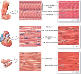
The term "striated muscle" refers to the striped appearance of skeletal and cardiac muscle under the microscope, due to the regular arrangement of contractile proteins. Smooth muscle lacks this pattern.
Neural Control of Muscle Contraction
All muscle types contract in response to electrical signals, but the source and pathway of these signals differ:
Skeletal muscle is controlled by the somatic nervous system (voluntary control).
Cardiac and smooth muscle are primarily regulated by the autonomic nervous system (involuntary control) and hormones.
Hearing and Sensory Processing in the Ear
Signal Transduction in Hair Cells
Auditory hair cells in the cochlea convert mechanical sound vibrations into electrical signals. The movement of the basilar membrane relative to the tectorial membrane bends the stereocilia on hair cells, opening or closing ion channels and altering neurotransmitter release. This process modulates the firing rate of sensory neurons, encoding sound information for the brain.
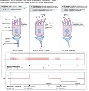
At rest: Some ion channels are open, and a baseline level of neurotransmitter is released.
Excitation (stereocilia bend toward the tallest): More channels open, increasing neurotransmitter release and action potentials in the sensory neuron.
Inhibition (stereocilia bend away): Channels close, reducing neurotransmitter release and action potentials.
Coding for Pitch and Loudness
The cochlea transforms sound waves into electrical signals. The basilar membrane varies in stiffness and width along its length, allowing different regions to resonate with different frequencies. The location of maximum movement encodes the pitch, while the magnitude of movement encodes loudness.
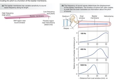
High-frequency sounds peak near the base (stiff region) of the basilar membrane.
Low-frequency sounds peak near the apex (flexible region).
Localization of Sound
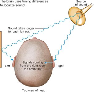
The brain determines the location of a sound source by comparing the timing and intensity of signals arriving at each ear. Unlike other senses, the auditory system does not use spatial receptive fields but relies on timing differences to compute location.
Types of Hearing Loss
Conductive hearing loss: Impaired transmission through the external or middle ear (often treatable with surgery).
Central hearing loss: Damage to neural pathways or the auditory cortex.
Sensorineural hearing loss: Damage to inner ear structures; hair cells cannot regenerate in mammals.
Skeletal Muscle Structure and Organization
Levels of Skeletal Muscle Organization
Skeletal muscle is composed of bundles of muscle fibers (cells), which are themselves composed of myofibrils. Each level is surrounded by connective tissue, and the entire muscle is richly supplied with blood vessels and nerves.
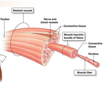
Muscle fiber: A single muscle cell, multinucleated, excitable, and capable of contraction.
Myofibril: A contractile element within the muscle fiber, composed of repeating units called sarcomeres.
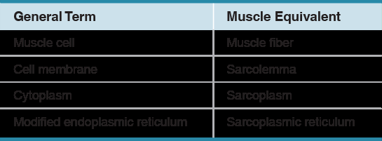
Muscle Fiber Terminology
Specialized terms are used for muscle cell structures:
General Term | Muscle Equivalent |
|---|---|
Muscle cell | Muscle fiber |
Cell membrane | Sarcolemma |
Cytoplasm | Sarcoplasm |
Modified endoplasmic reticulum | Sarcoplasmic reticulum |
T-Tubules and Sarcoplasmic Reticulum
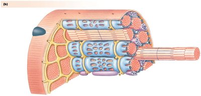
T-tubules are extensions of the sarcolemma that penetrate into the muscle fiber, allowing action potentials to rapidly reach the interior. The sarcoplasmic reticulum stores calcium ions, which are essential for muscle contraction.
Antagonistic Muscle Groups
Muscles often work in pairs called antagonistic muscle groups. When one muscle contracts (flexor), the other relaxes (extensor), allowing controlled movement of bones at joints.
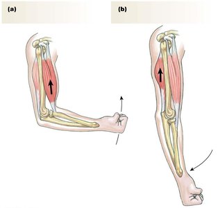
Flexor: Brings bones closer together (e.g., biceps).
Extensor: Moves bones farther apart (e.g., triceps).
Sarcomere Structure and Muscle Contraction
Sarcomere Organization
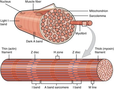
The sarcomere is the functional unit of muscle contraction, defined as the region between two Z discs. It contains interdigitated thick (myosin) and thin (actin) filaments. Sarcomeres shorten during contraction, pulling the Z discs closer together and shortening the muscle fiber.
Thick and Thin Filaments
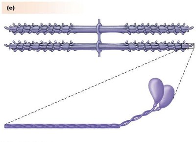
Thick filaments: Composed of myosin molecules, each with a head (binds actin and ATP) and a tail (forms the filament core).
Thin filaments: Composed of actin, tropomyosin, and troponin. Tropomyosin blocks myosin-binding sites on actin at rest; troponin moves tropomyosin in response to Ca2+.
Sliding Filament Theory and Cross-Bridge Cycle
Muscle contraction occurs as myosin heads bind to actin, pulling the thin filaments toward the center of the sarcomere. This process is powered by ATP and regulated by Ca2+ release from the sarcoplasmic reticulum.
At rest, myosin heads are cocked and weakly bound to actin; tropomyosin blocks strong binding.
Upon stimulation, Ca2+ binds troponin, shifting tropomyosin and exposing binding sites.
Myosin binds strongly, performs a power stroke, and slides actin filaments inward.
Key equation (ATP hydrolysis):
Summary: Muscle contraction is a highly organized process involving neural control, specialized muscle structures, and the coordinated action of contractile proteins. Understanding these mechanisms is essential for comprehending movement, force generation, and the integration of sensory and motor systems in the human body.
