 Back
BackMuscle Physiology: Mechanisms of Contraction, Metabolism, and Fiber Types
Study Guide - Smart Notes
 Tailored notes based on your materials, expanded with key definitions, examples, and context.
Muscle Physiology: Mechanisms of Contraction, Metabolism, and Fiber Types
Muscle Tension and Sarcomere Length
The amount of tension a muscle fiber can produce depends on the degree of overlap between thick and thin filaments within the sarcomere at the time of stimulation. This relationship is crucial for understanding how muscles generate force and why there is an optimal length for maximal tension production.
Optimal Sarcomere Length: Maximum tension is produced when the zone of overlap between thick and thin filaments is large, but the thin filaments do not extend across the sarcomere’s center.
Short Sarcomere Lengths: If the sarcomere is too short, thin filaments interfere with each other, reducing tension.
Overstretched Sarcomeres: If stretched too far, the zone of overlap is reduced, and cross-bridge interactions decrease, leading to diminished tension.
Extreme Stretching: When the zone of overlap is zero, no active tension can be produced. This is normally prevented by titin filaments and connective tissues.
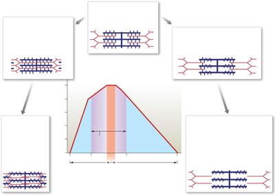
Key Point: The normal range of sarcomere lengths in the body is 75 to 130 percent of the optimal length.
Muscle Twitch and Phases of Contraction
A muscle twitch is the response of a muscle fiber to a single stimulus, consisting of three phases: latent period, contraction phase, and relaxation phase. The duration and characteristics of a twitch vary depending on the muscle type and external factors.
Latent Period: The brief delay between stimulus and contraction, during which the action potential propagates and calcium ions are released.
Contraction Phase: Tension rises to a peak as cross-bridge cycling occurs.
Relaxation Phase: Tension falls as calcium is reabsorbed and cross-bridges detach.
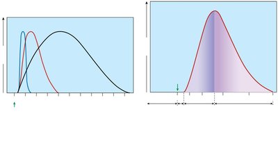
Example: The gastrocnemius muscle exhibits a clear latent period before contraction begins.
Types of Muscle Contraction: Treppe, Wave Summation, and Tetanus
Muscle fibers can respond to repeated stimuli in several ways, leading to different patterns of contraction and tension development.
Treppe: Successive stimuli delivered after the relaxation phase cause a gradual increase in peak tension due to increased calcium availability.
Wave Summation: Successive stimuli arrive before relaxation is complete, causing the effects of each twitch to add together.
Incomplete Tetanus: Stimulus frequency increases further, and tension rises to a peak with brief relaxation periods.
Complete Tetanus: Stimulus frequency is so high that relaxation is eliminated, and tension plateaus at a maximum level.
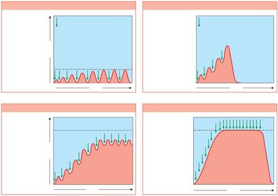
Key Point: Tetanus is achieved when the sarcoplasmic reticulum cannot reclaim calcium quickly enough, resulting in prolonged contraction.
Motor Units and Muscle Tone
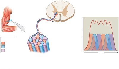
A motor unit consists of a motor neuron and all the muscle fibers it controls. The arrangement and activity of motor units determine the force and precision of muscle contractions.
Motor Unit Recruitment: Activation of additional motor units increases muscle tension. Recruitment starts with smaller, slower units and progresses to larger, more powerful ones.
Synchronous Motor Unit Summation: Motor units are activated on a rotating basis, allowing brief rest and recovery, which supports sustained contractions at slightly less than maximum tension.
Muscle Tone (Tonus): Continuous, low-level activity in motor units keeps muscles firm and ready for action, stabilizing limbs and increasing metabolism.
Types of Muscle Contractions: Isotonic and Isometric
Muscle contractions can be classified based on changes in muscle length and tension.
Isotonic Contraction: Tension remains constant while muscle length changes. Includes concentric (muscle shortens) and eccentric (muscle lengthens) contractions.
Isometric Contraction: Muscle length remains constant while tension increases.
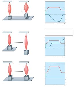
Example: Lifting a weight involves concentric contraction, while lowering it slowly involves eccentric contraction. Holding a weight steady involves isometric contraction.
Muscle Metabolism and Energy Sources
Muscle fibers require ATP for contraction, which is supplied by several energy sources depending on activity level.
ATP: Immediate energy source, but limited in quantity.
Creatine Phosphate (CP): Stores high-energy phosphate groups to rapidly regenerate ATP.
Glycogen: Provides glucose for glycolysis (anaerobic) and aerobic metabolism.
Energy Source | Utilization Process | Initial Available Quantity | Number of Twitches Supported | Duration of Isometric Tetanus Supported |
|---|---|---|---|---|
ATP | ATP → ADP + Pi | 3 mmol | 7 | 2 sec |
CP | ADP + CP → ATP + C | 30 mmol | 70 | 15 sec |
Glycogen (anaerobic) | Glycolysis | 100 mmol | 670 | 130 sec |
Glycogen (aerobic) | Aerobic metabolism | 100 mmol | 12,000 | 2400 sec (40 min) |

Key Point: During rest, muscles build up reserves of glycogen and CP. During moderate activity, aerobic metabolism predominates. During peak activity, glycolysis becomes the main source of ATP, leading to lactate accumulation and fatigue.
Muscle Fiber Types: Fast, Slow, and Intermediate
Muscle fibers are classified based on their contraction speed, fatigue resistance, and metabolic properties.
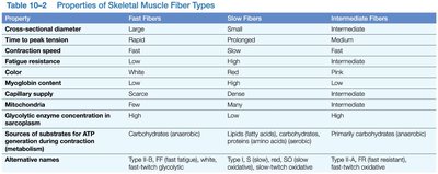
Fast Fibers (Type II-B): Large diameter, rapid contraction, low fatigue resistance, few mitochondria, pale color, rely on anaerobic metabolism.
Slow Fibers (Type I): Small diameter, slow contraction, high fatigue resistance, many mitochondria, dark color due to myoglobin, rely on aerobic metabolism.
Intermediate Fibers (Type II-A): Intermediate properties between fast and slow fibers.
Property | Fast Fibers | Slow Fibers | Intermediate Fibers |
|---|---|---|---|
Cross-sectional diameter | Large | Small | Intermediate |
Time to peak tension | Rapid | Prolonged | Medium |
Contraction speed | Fast | Slow | Fast |
Fatigue resistance | Low | High | Intermediate |
Color | White | Red | Pink |
Myoglobin content | Low | High | Low |
Capillary supply | Few | Many | More than fast, less than slow |
Mitochondria | Few | Many | Intermediate |
Glycolytic enzyme concentration | High | Low | High |
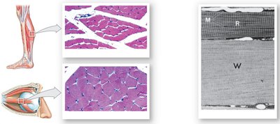
Example: Postural muscles contain more slow fibers, while muscles used for rapid, intense movements contain more fast fibers.
Muscle Hypertrophy and Atrophy
Muscle mass can increase (hypertrophy) or decrease (atrophy) depending on activity, nutrition, and innervation.
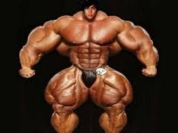
Hypertrophy: Enlargement of muscle due to repeated exhaustive stimulation, resulting in more mitochondria, myofibrils, glycogen reserves, and glycolytic enzymes.
Atrophy: Loss of muscle mass, tone, and power due to disuse, malnutrition, or loss of nerve supply.
Cardiac and Smooth Muscle Tissue
Cardiac and smooth muscle tissues have unique structural and functional characteristics compared to skeletal muscle.
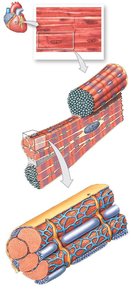
Cardiac Muscle
Found only in the heart
Uninucleate, striated, with intercalated discs
Short, broad T tubules; no terminal cisternae
Involuntary control
Smooth Muscle
Found in most organs and all blood vessels except capillaries
Uninucleate, non-striated, spindle-shaped cells
No thick and thin filament arrangement as in skeletal muscle
Uses calmodulin (not troponin) for calcium binding
Exhibits plasticity—can function over a wide range of lengths
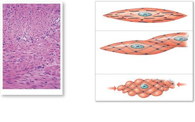
Key Point: Cardiac muscle is specialized for rhythmic contraction, while smooth muscle is adapted for slow, sustained contractions in various organs.
