 Back
BackMuscle Physiology: Structure and Function of Muscle Tissue
Study Guide - Smart Notes
 Tailored notes based on your materials, expanded with key definitions, examples, and context.
Muscle Tissue
Types of Muscle Tissue
Muscle tissue is specialized for contraction and is essential for movement, stability, and various physiological functions. There are three main types of muscle tissue: skeletal, cardiac, and smooth muscle.
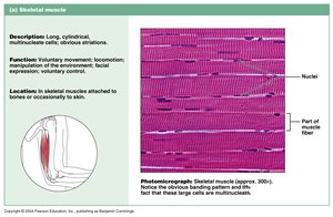
Skeletal Muscle: Voluntary, striated muscle attached to bones; responsible for body movement.
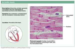
Cardiac Muscle: Involuntary, striated muscle found only in the heart; responsible for pumping blood.
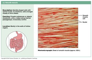
Smooth Muscle: Involuntary, non-striated muscle found in walls of hollow organs; responsible for propelling substances.
Functions of Muscle Tissue
Muscle tissue performs several critical functions in the body:
Movement: Muscles contract to produce movement of the body and its parts.
Opening and Closing: Muscles regulate passageways and openings (e.g., sphincters).
Stabilizing and Containing: Muscles stabilize joints and maintain posture.
Temperature Control: Muscle contraction generates heat, accounting for approximately 85% of body heat.
Muscle Characteristics
Properties of Muscle Tissue
Muscle tissue exhibits four key characteristics:
Excitability: Ability to respond to stimuli and produce electrical signals.
Contractility: Ability to shorten forcibly when stimulated.
Extensibility: Ability to stretch without being damaged.
Elasticity: Ability to return to original shape after contraction or extension.
Muscle Organization
Functional Groups of Muscles
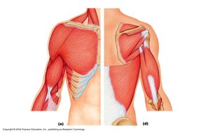
Muscles are organized into functional groups that perform similar actions. Each group consists of individual muscles attached to bones by tendons.
Example: Biceps, brachialis, and triceps are functional groups involved in arm movement.
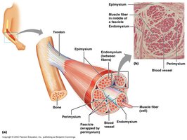
Muscle Structure: Fascicles and Wrappings
Muscles are composed of bundles called fascicles, which are groups of muscle fibers (cells). Several connective tissue layers surround and organize muscle components:
Deep Fascia: Surrounds functional groups and attaches to bone or tendons.
Epimysium: Surrounds individual muscles.
Perimysium: Surrounds fascicles.
Endomysium: Surrounds individual muscle cells.
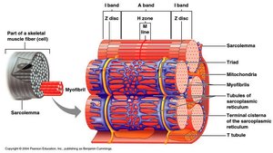
Microscopic Anatomy of Muscle
Muscle Cell Structure
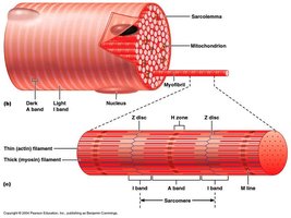
Muscle cells (fibers) have specialized structures for contraction:
Sarcolemma: Cell membrane of muscle fiber.
Sarcoplasmic Reticulum: Specialized endoplasmic reticulum that stores calcium ions (Ca++).
Myofibrils: Cylindrical structures within muscle fibers, composed of myofilaments.

Myofilaments
Myofibrils contain three types of myofilaments:
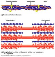
Thin Filaments: Composed primarily of actin, with associated proteins tropomyosin and troponin.
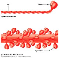
Thick Filaments: Composed of myosin molecules, each with two heads and a tail.
Elastic Filaments: Composed of titin, which anchors thick filaments to the Z disc.

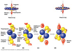
Structure of Thin and Thick Filaments
Thin filaments consist of actin molecules with myosin binding sites, tropomyosin (blocks binding sites), and troponin (moves tropomyosin). Thick filaments are made of myosin molecules with heads that bind to actin.
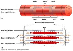
Sarcomere Structure
The sarcomere is the functional unit of muscle contraction, defined as the region between two Z discs. It contains overlapping thick and thin filaments, creating distinct bands:
A Band: Length of thick filament, including overlap with thin filaments.
H Zone: Region with only thick filaments.
I Band: Region with only thin filaments.
Z Disc: Boundary of the sarcomere.
M Line: Center of the sarcomere.
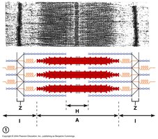
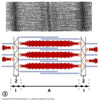
Sarcomere Changes During Contraction
During muscle contraction, actin filaments are drawn together, causing the H zone and I band to disappear, Z discs move closer, and the A band remains unchanged.
Muscle Contraction Mechanism
Steps in Muscle Contraction
Muscle contraction is initiated by a motor neuron:
Motor neuron fires, sending an electrical signal.
Signal travels down the neuron and stimulates the muscle cell.
Electrical signal spreads along the sarcolemma and into T tubules.
Sarcoplasmic reticulum releases Ca++.
Ca++ binds to troponin, triggering contraction.
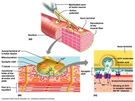
Neuromuscular Junction
The neuromuscular junction is where the motor neuron meets the muscle cell. Key structures include:
Axon Terminal: End of the neuron.
Synaptic End Bulbs: Contain synaptic vesicles filled with acetylcholine (Ach).
Motor Endplate: Region of muscle cell membrane with Ach receptors.

Stimulation of Muscle
Sequence of events at the neuromuscular junction:
Electrical signal reaches synaptic end bulbs.
Ca++ enters end bulbs, causing vesicles to release Ach.
Ach diffuses across synaptic cleft and binds to receptors on sarcolemma.
Ion gates open, generating a new electrical signal in the muscle cell.
Sarcoplasmic reticulum releases Ca++.
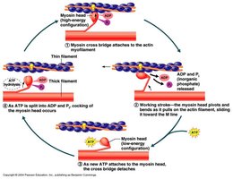
Sliding Filament Theory
Muscle contraction occurs via the sliding filament mechanism:
Ca++ binds to troponin, moving tropomyosin and exposing myosin binding sites on actin.
ATP activates myosin, allowing it to bind to actin.
Myosin pulls actin, causing contraction.
ATP binds to myosin, causing it to release actin and repeat the cycle.

Recovery After Contraction
After contraction, acetylcholinesterase breaks down Ach, Ca++ is actively transported back into the sarcoplasmic reticulum, and filaments return to resting position.
Motor Units and Muscle Contraction Strength
Motor Neurons and Motor Units
A motor unit consists of a motor neuron and all the muscle fibers it innervates. The number of motor units activated determines the strength of contraction.
Fine Movements: Many small motor units (e.g., fingers, eyes).
Strong Movements: Fewer, larger motor units (e.g., back, legs).
Summary Table: Muscle Types
Muscle Type | Structure | Function | Location |
|---|---|---|---|
Skeletal | Long, cylindrical, multinucleate, striated | Voluntary movement | Attached to bones |
Cardiac | Branching, striated, intercalated discs | Pumps blood | Heart walls |
Smooth | Spindle-shaped, non-striated | Propels substances | Walls of hollow organs |
Key Equations
Muscle contraction strength is proportional to the number of motor units activated:
Strength of Contraction:
Additional info:
Muscle contraction is regulated by calcium ions and ATP, and involves complex interactions between actin and myosin filaments.
Motor units allow graded control of muscle contraction, enabling both fine and powerful movements.
