 Back
BackMuscle Physiology: Types, Structure, and Control
Study Guide - Smart Notes
 Tailored notes based on your materials, expanded with key definitions, examples, and context.
Muscle Tissue Types and Their Characteristics
Overview of Muscle Tissue
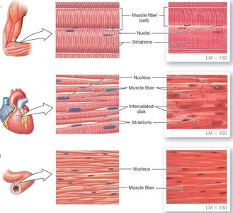
Muscle tissue is essential for movement and force generation in the human body. There are three main types of muscle tissue, each with distinct structural and functional properties.
Skeletal Muscle: Large, multinucleated cells; striated appearance; attached to bones; responsible for voluntary movements.
Cardiac Muscle: Smaller, branched cells with one nucleus; striated; found only in the heart; responsible for pumping blood.
Smooth Muscle: Small, spindle-shaped cells; not striated; found in internal organs; controls involuntary movements of materials within the body.
Striated muscle refers to muscle tissue with a striped appearance under the microscope, due to the arrangement of contractile proteins. Both skeletal and cardiac muscles are considered striated.
Control of Muscle Contraction
Neural and Endocrine Regulation
Muscle contraction is initiated by signals from the nervous system, and in some cases, the endocrine system. The type of control depends on the muscle tissue:
Skeletal Muscle: Controlled by the somatic nervous system (voluntary); a single motor neuron directly innervates each muscle fiber.
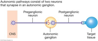
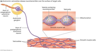
Cardiac and Smooth Muscle: Controlled by the autonomic nervous system (involuntary) and hormones; involves a two-neuron pathway with synapses in autonomic ganglia.
All muscle types initiate contraction in response to an action potential (electrical signal).
Skeletal Muscle Structure
Organization of Skeletal Muscle
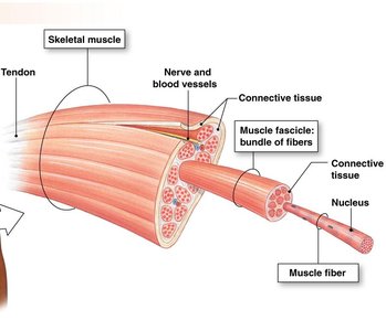
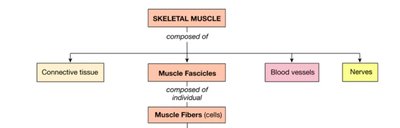
Skeletal muscle is composed of bundles of muscle fibers (cells), which are grouped together and encased in connective tissue. Each muscle fiber is a fused cell with multiple nuclei and is described using the "sarco-" prefix (e.g., sarcolemma, sarcoplasm).
Muscle Fiber: The basic unit; excitable and capable of contraction.
Muscle Fascicle: Bundle of muscle fibers surrounded by connective tissue.
Skeletal Muscle: Composed of multiple fascicles, connective tissue, blood vessels, and nerves.
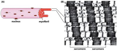
Striated Pattern and Sarcomeres
The striated appearance of skeletal muscle arises from the arrangement of contractile proteins within sarcomeres, the functional units of muscle fibers. Sarcomeres are composed of actin and myosin filaments, which slide past each other during contraction.
Sarcomere: The repeating unit within a myofibril; responsible for muscle contraction.
Myofibril: Bundle of sarcomeres within a muscle fiber.
Example: When an action potential reaches a muscle fiber, calcium ions are released, triggering the sliding of actin and myosin filaments and resulting in muscle shortening.
Antagonistic Muscle Groups
Definition and Function
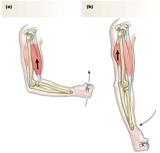
Antagonistic muscle groups are pairs of muscles that produce opposite movements at a joint. One muscle contracts to produce flexion, while the other contracts to produce extension.
Flexor: Brings the centers of connected bones closer together when contracted.
Extensor: Moves bones further apart when contracted.
Example: The biceps and triceps in the arm are antagonistic muscles; the biceps flexes the elbow, while the triceps extends it.
Summary Table: Muscle Tissue Types
The following table summarizes the main characteristics of the three muscle tissue types:
Muscle Type | Cell Structure | Striated? | Location | Control |
|---|---|---|---|---|
Skeletal | Large, multinucleated | Yes | Attached to bones | Somatic (voluntary) |
Cardiac | Small, branched, one nucleus | Yes | Heart | Autonomic (involuntary) |
Smooth | Small, spindle-shaped, one nucleus | No | Internal organs | Autonomic (involuntary) |
Additional info:
The "sarco-" prefix is used for muscle cell components (e.g., sarcolemma = muscle cell membrane).
Muscle contraction is triggered by an action potential, which leads to calcium release and filament sliding.
Antagonistic muscle groups are essential for coordinated movement and joint stability.
