 Back
BackMuscle Terminology and Classification: Structure, Naming, and Function
Study Guide - Smart Notes
 Tailored notes based on your materials, expanded with key definitions, examples, and context.
Muscle Terminology and Classification
Muscle Roles in Movement
Muscles are classified based on their roles during movement. Understanding these terms is essential for describing muscle function in anatomy and physiology.
Prime mover (agonist): The muscle primarily responsible for producing a specific movement.
Antagonist: The muscle that opposes the action of the agonist.
Synergist: A muscle that assists the agonist in performing its action.
Fixator: A muscle that stabilizes the origin of the agonist so that it can act efficiently.
Muscle Naming Conventions
Muscles are named according to several criteria, which help identify their location, structure, and function.
Location: Muscles are often named for the region of the body where they are found (e.g., deltoid in the shoulder, brachialis in the arm).
Origin and Insertion: Some muscles are named for their points of attachment (e.g., sternocleidomastoid originates at the sternum and clavicle and inserts at the mastoid process).
Number of Origins: Muscles may be named for the number of tendons of origin (e.g., biceps brachii has two origins, triceps brachii has three).
Direction of Muscle Fibers: The orientation of fibers can be reflected in the name (e.g., rectus abdominis runs straight, transversus abdominis runs transversely).
Shape: Muscles may be named for their shape (e.g., deltoid is triangular, trapezius is trapezoidal).
Relative Size: Terms like maximus (largest), minimus (smallest), longus (long), and brevis (short) indicate size.
Action: The function of the muscle may be included in its name (e.g., flexor carpi radialis flexes the wrist).
Historical References: Some muscles are named based on historical or descriptive references.
Examples of Muscle Naming Criteria
Location: Temporalis (temporal region of the skull), pectoralis major (chest).
Origin/Insertion: Sternocleidomastoid (sternum, clavicle, mastoid process).
Number of Origins: Biceps brachii (two origins), triceps brachii (three origins).
Direction of Fibers: Rectus abdominis (straight), oblique (angled).
Shape: Deltoid (delta-shaped), trapezius (trapezoid-shaped).
Size: Gluteus maximus (largest gluteal muscle), gluteus minimus (smallest gluteal muscle).
Action: Flexor digitorum longus (flexes the toes).
Muscle Anatomy: Major Groups and Examples
The human body contains numerous muscles, each with specific functions and anatomical locations. Below are some major muscle groups and examples:
Head and Neck: Temporalis, masseter, sternocleidomastoid, platysma
Thorax: Pectoralis major, pectoralis minor, serratus anterior, intercostals
Abdomen: Rectus abdominis, external oblique, internal oblique, transversus abdominis
Shoulder and Arm: Deltoid, trapezius, biceps brachii, triceps brachii, brachialis
Forearm: Brachioradialis, flexor carpi radialis, extensor carpi ulnaris
Pelvis and Thigh: Iliopsoas, pectineus, rectus femoris, vastus lateralis, adductor longus
Leg: Gastrocnemius, soleus, fibularis longus, tibialis anterior
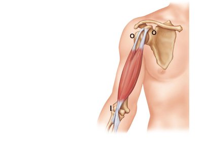
Muscle Attachments: Origin and Insertion
Muscles attach to bones at two points: the origin and the insertion. The origin is typically the fixed attachment, while the insertion moves with contraction.
Origin (O): The stationary attachment point of the muscle.
Insertion (I): The movable attachment point.
Example: The biceps brachii originates at the scapula and inserts at the radius, allowing flexion of the forearm.
Muscle Fiber Arrangement and Function
The arrangement of muscle fibers affects the muscle's range of motion and power. Common arrangements include parallel, pennate, convergent, and circular.
Parallel: Fibers run parallel to the long axis (e.g., sartorius).
Pennate: Fibers attach obliquely to a central tendon (e.g., rectus femoris).
Convergent: Fibers converge from a broad origin to a single insertion (e.g., pectoralis major).
Circular: Fibers arranged in concentric rings (e.g., orbicularis oris).
Summary Table: Muscle Naming Criteria
Criterion | Example | Description |
|---|---|---|
Location | Temporalis | Located in the temporal region |
Origin/Insertion | Sternocleidomastoid | Originates at sternum/clavicle, inserts at mastoid |
Number of Origins | Biceps brachii | Two origins |
Direction of Fibers | Rectus abdominis | Fibers run straight |
Shape | Deltoid | Delta-shaped |
Size | Gluteus maximus | Largest gluteal muscle |
Action | Flexor digitorum longus | Flexes the toes |
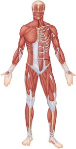
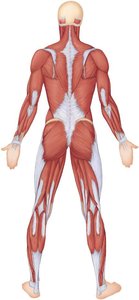
Major Muscles of the Human Body
The following images illustrate the major muscle groups of the human body, highlighting their anatomical locations and relationships.
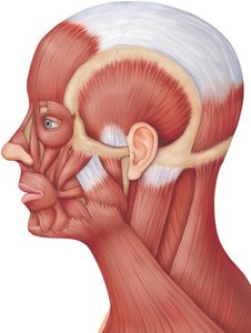
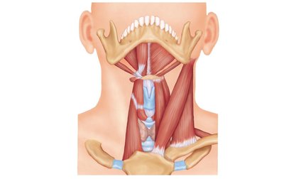
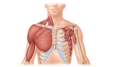
Muscle Groups of the Head, Neck, and Thorax
Muscles in these regions are responsible for facial expression, mastication, head movement, and respiration.
Head: Epicranius, orbicularis oculi, zygomaticus, orbicularis oris
Neck: Sternohyoid, sternocleidomastoid, platysma
Thorax: Pectoralis major, pectoralis minor, serratus anterior, intercostals
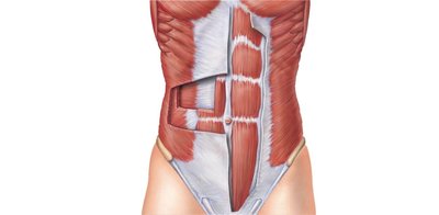
Muscles of the Abdominal Wall
The abdominal wall muscles support the trunk, allow movement, and help maintain posture.
Rectus abdominis: Flexes the vertebral column.
External oblique: Compresses the abdomen and flexes the spine.
Internal oblique: Assists in trunk rotation and lateral flexion.
Transversus abdominis: Compresses abdominal contents.
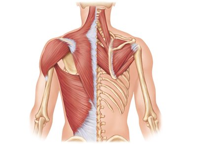
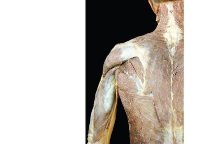
Muscles of the Shoulder and Arm
These muscles facilitate movement of the shoulder and arm, including abduction, flexion, and extension.
Deltoid: Abducts the arm.
Trapezius: Elevates, retracts, and rotates the scapula.
Biceps brachii: Flexes the forearm.
Triceps brachii: Extends the forearm.
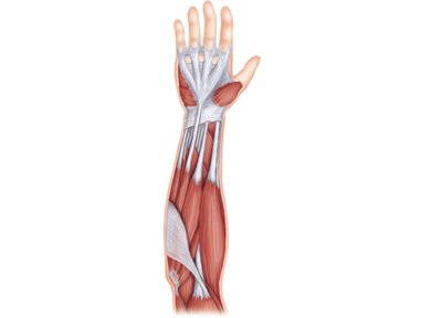
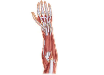
Muscles of the Forearm and Hand
Forearm muscles control movements of the wrist, hand, and fingers, including flexion, extension, pronation, and supination.
Flexor carpi radialis: Flexes and abducts the wrist.
Extensor carpi ulnaris: Extends and adducts the wrist.
Pronator teres: Pronates the forearm.
Brachioradialis: Flexes the forearm.
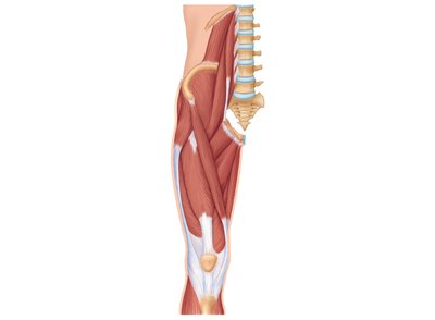
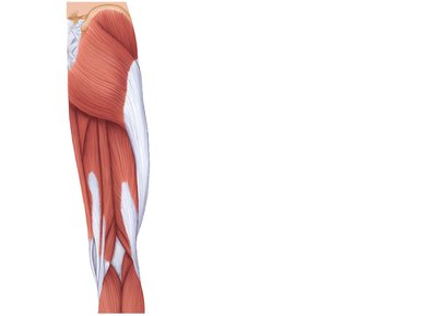
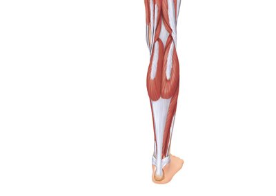
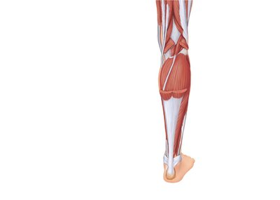
Muscles of the Thigh and Leg
These muscles are essential for locomotion, supporting body weight, and maintaining balance.
Quadriceps femoris: Extends the knee.
Hamstrings: Flex the knee and extend the hip.
Gastrocnemius: Plantar flexes the foot.
Soleus: Plantar flexes the foot.
Tibialis anterior: Dorsiflexes the foot.
Summary
Muscle terminology and classification are foundational for understanding human anatomy and physiology. Muscles are named based on their location, structure, and function, and their arrangement determines their mechanical properties. Knowledge of major muscle groups and their actions is essential for clinical and academic applications.
Additional info: Academic context was added to expand brief points and clarify muscle naming conventions, fiber arrangements, and functional roles.
