 Back
BackMuscle Tissue and Muscle Cell Structure: An Overview
Study Guide - Smart Notes
 Tailored notes based on your materials, expanded with key definitions, examples, and context.
Muscle Tissue: Types and General Structure
Overview of Muscle Tissue
Muscle tissue is a specialized tissue found throughout the body, responsible for producing movement, maintaining posture, and generating heat. Muscle cells, also known as myocytes, are the fundamental units of muscle tissue and are surrounded by an extracellular matrix called the endomysium, which helps hold the cells together within the tissue.
Types of Muscle Tissue
Striated Muscle: Characterized by alternating light and dark bands (striations). Includes skeletal muscle and cardiac muscle.
Smooth Muscle: Lacks striations and appears smooth under the microscope. Found in the walls of hollow organs, eyes, skin, and certain ducts.

Skeletal Muscle Tissue
Structure and Function
Skeletal muscle tissue is primarily responsible for voluntary movements and is attached to bones via connective tissue. The muscle fibers are long, multinucleated, and arranged in parallel. Each fiber contains multiple nuclei due to the fusion of embryonic cells called myoblasts, and numerous mitochondria to supply ATP for contraction.
Striated fibers are named for their length and banded appearance.
Voluntary control is exerted by conscious thought.

Cardiac Muscle Tissue
Structure and Function
Cardiac muscle tissue forms the heart and is responsible for pumping blood. The cells are short, wide, branched, and typically have a single nucleus. They are striated and connected by intercalated discs, which facilitate synchronized contraction. Cardiac muscle operates involuntarily, under unconscious control.
Smooth Muscle Tissue
Structure and Function
Smooth muscle tissue lines hollow organs and structures. The myocytes are long, flattened, and have two pointed ends with a single, centrally located nucleus. These cells are linked by gap junctions and contract involuntarily. Smooth muscle lacks striations, giving it a uniform appearance.

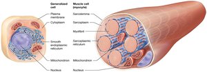
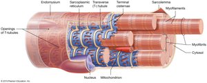
Muscle Cell Structure
Specialized Organelles and Terminology
Muscle cells share similarities with generalized animal cells but have specialized names for certain structures:
Sarcoplasm: Cytoplasm of a muscle cell
Sarcolemma: Plasma membrane of a muscle cell
Myofibrils: Cylindrical organelles containing bundles of specialized proteins necessary for contraction
Sarcoplasmic reticulum: Modified endoplasmic reticulum, forms a web-like network around each myofibril
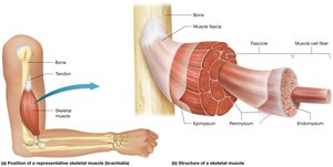
Connective Tissue Layers in Skeletal Muscle
Organization and Function
Skeletal muscle is organized into several layers of connective tissue:
Epimysium: Outermost layer, surrounds the entire muscle
Perimysium: Surrounds bundles of muscle fibers called fascicles
Endomysium: Surrounds each individual muscle fiber
The fascia separates individual muscles and forms tendons and aponeuroses, connecting muscles to bones and to other muscles.
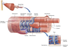
Structures of Skeletal Muscle Fibers
Transverse Tubules and Triads
Transverse tubules (T-tubules) are extensions of the sarcolemma that tunnel into the muscle fiber, surrounding each myofibril and filled with extracellular fluid. The terminal cisternae are enlarged portions of the sarcoplasmic reticulum that, together with a T-tubule, form a triad. This structure is essential for the rapid transmission of action potentials and the release of calcium ions during muscle contraction.

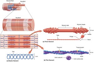
Myofibril Structure and Myofilaments
Types of Myofilaments
Myofibrils are composed of three main types of myofilaments:
Thick filaments: Made of myosin, a contractile protein with two globular heads and two intertwining tails. The heads bind to actin on the thin filaments.
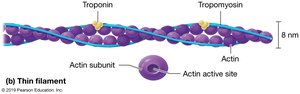
Thin filaments: Composed of actin (contractile protein), tropomyosin (regulatory protein that covers active sites on actin), and troponin (regulatory protein that holds tropomyosin in place).
Elastic filaments: Made of titin, a structural protein that provides elasticity and stabilizes the thick filaments.

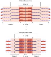
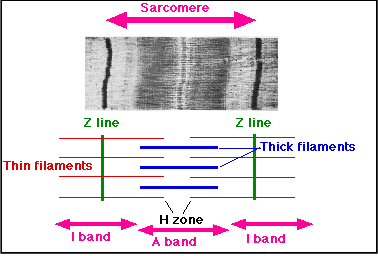
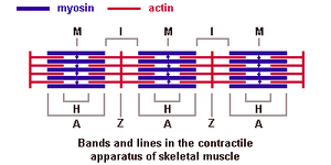
Myofilament Arrangement and Sarcomere Structure
Bands, Lines, and Functional Units
The arrangement of myofilaments creates distinct bands and lines within the myofibril:
I Bands: Lighter regions containing only thin filaments
A Bands: Dark regions containing thick filaments
H Zone: Slightly lighter area in the middle of the A band, contains only thick filaments
M Line: Dark line in the center of the A band, holds thick filaments in place
Z-Disc: Dark line in the I band, anchors thin filaments and marks the boundary of a sarcomere
The sarcomere is the functional unit of muscle contraction, spanning from one Z-disc to the next. During contraction, actin filaments slide past myosin filaments, shortening the sarcomere.

Cardiac and Smooth Muscle Cell Structure
Cardiac Muscle Cells
Cardiac muscle cells are striated, contain sarcomeres, and have T-tubules and an extensive sarcoplasmic reticulum. Intercalated discs connect the cells, allowing for coordinated contraction.
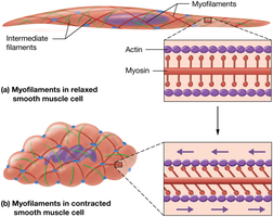
Smooth Muscle Cells
Smooth muscle cells lack striations, have more thin filaments than thick, and do not contain troponin. The filaments are arranged obliquely and anchored to dense bodies. T-tubules are absent, and the sarcoplasmic reticulum is less extensive.
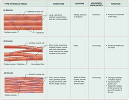
Summary Table: Types of Muscle Tissue
Type of Muscle Tissue | Structure | Location | Voluntary/Involuntary | Function |
|---|---|---|---|---|
Skeletal | Long, cylindrical, multinucleated, striated | Mostly attached to skeleton | Voluntary | Produces movement of the body |
Cardiac | Short, wide, branching, single nucleus, striated, intercalated discs | Heart | Involuntary | Pumps blood through the heart |
Smooth | Long, flattened, single nucleus, non-striated | Walls of hollow organs, eyes, skin, ducts | Involuntary | Changes diameter of tubes, moves substances |
Key Terms and Concepts
Myocyte: Muscle cell
Myofibril: Organelle within muscle cell, composed of myofilaments
Myofilament: Protein filament (thick, thin, elastic) responsible for contraction
Sarcomere: Functional unit of contraction, defined by Z-discs
Fascia: Connective tissue separating muscles
Tendon: Connects muscle to bone
Aponeurosis: Connects muscle to muscle
Summary of Muscle Contraction
During muscle contraction, the thin filaments (actin) slide past the thick filaments (myosin), causing the sarcomere to shorten. This process is fundamental to all types of muscle tissue, though the structural details vary between skeletal, cardiac, and smooth muscle.
