 Back
Muscle Tissue and Muscle Contraction Physiology: Structured Study Notes
Study Guide - Smart Notes
 Tailored notes based on your materials, expanded with key definitions, examples, and context.
Muscle Tissue Types
Overview of Muscle Tissue
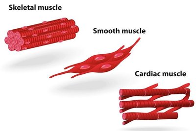
Muscle tissue is essential for movement and force generation in the human body. There are three main types of muscle tissue, each with distinct structural and functional characteristics.
Skeletal muscle: Attached to bones, striated, voluntary control.
Cardiac muscle: Makes up the heart wall, striated, involuntary control.
Smooth muscle: Found in internal organs, non-striated, involuntary control.
Features and Structure of Skeletal Muscle
Organization and Components
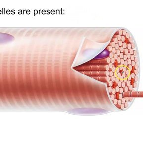
Skeletal muscle is composed of bundles organized hierarchically to facilitate movement. It is under conscious control and considered an organ due to its multiple tissue types.
Muscle fiber: A single muscle cell, long and thin, contains myofibrils.
Myofibrils: Rod-like structures running the length of the cell, organized in sarcomeres.
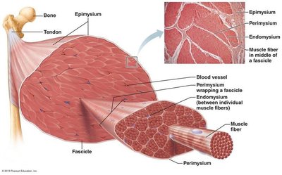
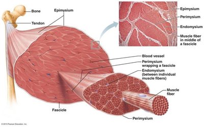
Fascicle: Bundle of muscle fibers wrapped in connective tissue.
Muscle: Bundle of fascicles wrapped in connective tissue, contains blood vessels and nerves.
Connective Tissue Layers
Connective tissue surrounds and organizes muscle components, providing support and facilitating force transmission.
Endomysium: Surrounds individual muscle fibers.
Perimysium: Surrounds fascicles.
Epimysium: Surrounds the entire muscle, beneath fascia.
Muscle Attachments
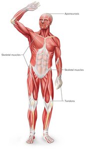
Muscles attach to bones and other structures via specialized connective tissues.
Tendon: Cord-like connective tissue connecting muscle to bone.
Aponeurosis: Sheet-like connective tissue connecting muscle to bone, skin, or other muscles.
Fascia: Thin connective tissue network supporting organs, muscles, bones, nerves, and blood vessels.
Muscle Cell (Fiber) Structure
Special Features of Muscle Cells
Muscle fibers are specialized cells with unique structures for contraction.
Multinucleated: Multiple nuclei per cell.
Sarcolemma: Cell membrane of muscle fiber.
Sarcoplasm: Cytoplasm of muscle fiber.
Myofibrils: Fill most of cytoplasm, organized in sarcomeres.
Sarcoplasmic reticulum: Specialized smooth ER storing calcium.
Myofibrils and Sarcomeres
Organization and Function
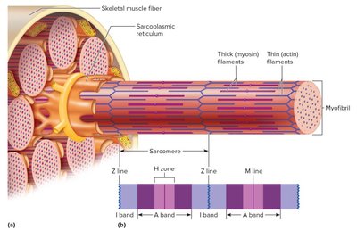
Myofibrils are divided into repeating units called sarcomeres, which are the fundamental contractile units of muscle.
Sarcomere: Comprised of actin (thin) and myosin (thick) filaments.
Sarcoplasmic reticulum: Stores calcium, essential for contraction.
T tubules: Carry electrical impulses to the sarcoplasmic reticulum.
Triad: Consists of one T tubule and two SR cisternae.
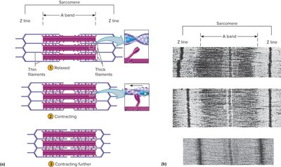
Sarcomere Structure
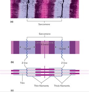
The sarcomere's arrangement of filaments gives skeletal muscle its striated appearance and defines its contractile properties.
I band: Light band, thin actin filaments.
A band: Dark band, thick myosin filaments (with overlap).
H zone: Center of A band, only thick filaments.
Z line: Sarcomere boundary, anchors filaments.
M line: Center of sarcomere, anchors thick filaments.
Sliding Filament Model of Contraction
Mechanism of Muscle Contraction
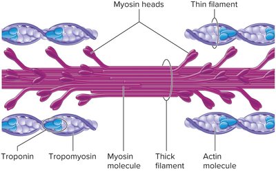
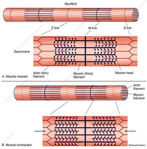
Muscle contraction occurs when myosin heads form cross-bridges with actin, pulling the filaments and shortening the sarcomere.
Myosin heads pull actin filaments toward the M-line.
H zones and I bands narrow, Z lines move closer.
Filament lengths remain unchanged; overlap increases.
Regulatory Proteins: Troponin and Tropomyosin
Role in Muscle Contraction
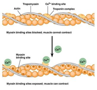
Troponin and tropomyosin regulate the formation of cross-bridges between actin and myosin, controlling muscle contraction.
Tropomyosin: Covers myosin binding sites on actin.
Troponin: Binds calcium, shifts tropomyosin to expose binding sites.
Calcium binding to troponin initiates contraction.
Cross-Bridge Cycling
Steps and Requirements
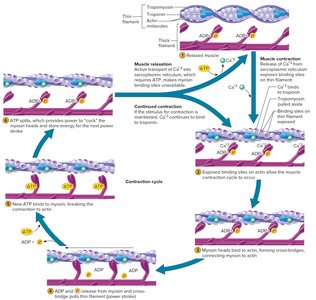
Cross-bridge cycling is the repeated process of myosin binding to actin, performing a power stroke, and detaching, which drives muscle contraction.
Requires calcium and ATP.
Steps: Calcium binds to troponin, myosin attaches to actin, power stroke, ATP binds to myosin, myosin detaches, ATP hydrolyzed to cock myosin head.
Muscle Contraction and Force Generation
From Sarcomere to Whole Muscle
As sarcomeres contract, the shortening is transmitted through myofibrils, muscle fibers, and the entire muscle, resulting in movement.
Force is transferred to bone via tendons.
Coordinated contraction creates meaningful movement.
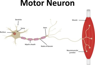
Excitation of Muscle: Neuromuscular Junction
Motor Neuron and Synapse
Muscle contraction is initiated by signals from motor neurons at the neuromuscular junction, a specialized synapse.
Motor neuron: Sends contraction signal to muscle.
Neuromuscular junction: Site of communication between neuron and muscle fiber.
Synapse: Where neurotransmitter is released.
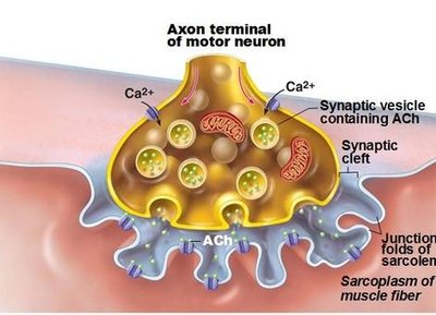
Parts of the Neuromuscular Junction
The neuromuscular junction consists of several specialized structures for efficient signal transmission.
Motor end plate: Folded region of muscle fiber membrane.
Synaptic cleft: Space between neuron and muscle fiber.
Synaptic vesicles: Contain neurotransmitter (acetylcholine).
Acetylcholine (Ach): Main neurotransmitter for skeletal muscle.
Excitation-Contraction Coupling (ECC)
Sequence of Events
ECC describes the process by which a neural signal leads to muscle contraction.
Motor neuron releases Ach at the neuromuscular junction.
Ach binds to sarcolemma receptors, generating an action potential.
Action potential travels along sarcolemma and T-tubule.
Calcium channels open, Ca2+ released from SR.
Ca2+ binds to troponin, tropomyosin shifts, myosin binds actin.
Power stroke occurs, contraction proceeds.
Ca2+ pumped back into SR after contraction.
Muscle Relaxation
Mechanisms of Relaxation
Muscle relaxation occurs when neural stimulation ceases and calcium is removed from the cytoplasm.
Acetylcholinesterase breaks down Ach in the synaptic cleft.
Calcium pump uses ATP to return Ca2+ to SR.
Troponin-tropomyosin complex covers actin binding sites.
Smooth Muscle
Structure and Function
Smooth muscle fibers are shorter, single-nucleated, and lack striations. They are found in internal organs and contract involuntarily.
Myofilaments randomly organized, no sarcomeres.
No transverse tubules, less developed SR.
Contraction involves actin and myosin, calcium binds to calmodulin.
Neurotransmitters: Acetylcholine and norepinephrine.
Cardiac Muscle
Structure and Function
Cardiac muscle is found only in the heart, is striated, and contracts in a linear fashion similar to skeletal muscle. Fibers branch and connect via intercalated discs.
Single nucleus per cell.
Network contracts as a unit (syncytium).
Self-exciting and rhythmic.
Clinical Applications: Muscular System Disorders
Myasthenia Gravis (MG)
MG is an autoimmune disorder where antibodies attack acetylcholine receptors, causing muscle weakness and fatigue.
Treatments: Acetylcholinesterase inhibitors, immunosuppressants, antibody therapy, plasma exchange.
Clostridium Botulinum
This bacterium produces a toxin that blocks acetylcholine release, causing muscle paralysis and symptoms in digestive, muscular, and respiratory systems.
Treatment: Antitoxin.
Medical use: Botox for cosmetic and therapeutic purposes.
Muscle Type | Striations | Control | Location | Special Features |
|---|---|---|---|---|
Skeletal | Yes | Voluntary | Attached to bones | Multinucleated, organized in sarcomeres |
Cardiac | Yes | Involuntary | Heart | Intercalated discs, syncytium |
Smooth | No | Involuntary | Internal organs | Single nucleus, calmodulin-mediated contraction |
Key Formula:
ATP hydrolysis for muscle contraction:
Calcium binding:
Excitation-Contraction Coupling:
