 Back
BackMuscle Tissue & Muscle Fibers: Structure, Types, and Function
Study Guide - Smart Notes
 Tailored notes based on your materials, expanded with key definitions, examples, and context.
Muscle Tissue Types and Their Characteristics
Overview of Muscle Tissue
Muscle tissue is specialized for contraction and is essential for movement, posture, and various involuntary processes in the body. There are three main types of muscle tissue: skeletal muscle, cardiac muscle, and smooth muscle. Each type has distinct structural and functional characteristics.
Main features | Location | Type of cells | Histology |
|---|---|---|---|
Fibers: striated, tubular, and multinucleated Voluntary Usually attached to skeleton | Skeleton | Long, cylindrical, multinucleated | Striated appearance |
Fibers: non-striated, spindle-shaped, and uninucleated Involuntary Usually covering walls of internal organs | Walls of hollow organs (e.g., intestines, blood vessels) | Spindle-shaped, uninucleated | Non-striated appearance |
Fibers: striated, branched, and uninucleated Involuntary Only covering walls of the heart | Heart | Branched, usually single nucleus | Striated with intercalated discs |
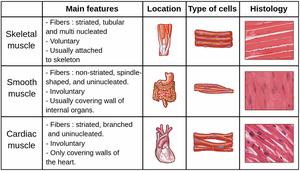
Skeletal Muscle Tissue
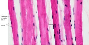
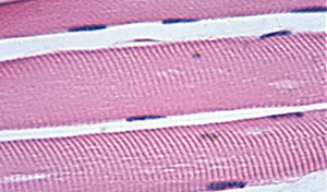
Skeletal muscle tissue consists of long, cylindrical fibers that are multinucleated and display prominent striations. These muscles are under voluntary control and are primarily responsible for body movement and posture.
Striations: Alternating light and dark bands due to the arrangement of actin and myosin filaments.
Nuclei: Multiple nuclei located at the periphery of the cell.
Location: Attached to bones via tendons.
Cardiac Muscle Tissue
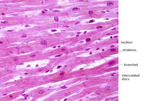
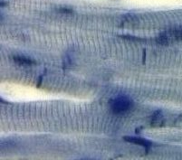
Cardiac muscle tissue is found only in the heart. Its fibers are striated, branched, and typically have a single central nucleus. Cardiac muscle is involuntary and features unique structures called intercalated discs that facilitate synchronized contraction.
Striations: Present, similar to skeletal muscle but less distinct.
Intercalated discs: Specialized junctions that connect cardiac muscle cells and allow rapid transmission of electrical impulses.
Branched fibers: Allow for a networked structure, supporting coordinated contractions.
Smooth Muscle Tissue
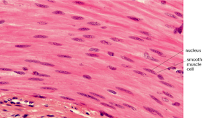
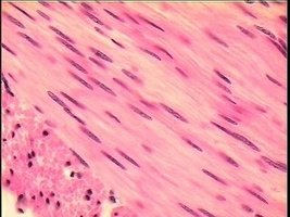
Smooth muscle tissue is composed of spindle-shaped, non-striated cells with a single central nucleus. It is found in the walls of hollow organs and is responsible for involuntary movements such as peristalsis and vasoconstriction.
No striations: The actin and myosin filaments are not arranged in a regular pattern.
Location: Walls of blood vessels, digestive tract, respiratory tract, urinary bladder, and uterus.
Function: Controls slow, sustained, involuntary movements.
Structure and Organization of Skeletal Muscle
Hierarchical Organization
Skeletal muscle is organized into several levels, from the whole muscle down to the molecular components responsible for contraction. This organization allows for efficient force generation and transmission.
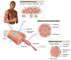
Muscle (organ): Surrounded by epimysium, contains bundles of fascicles.
Fascicle: Bundle of muscle fibers, surrounded by perimysium.
Muscle fiber (cell): Surrounded by endomysium, contains myofibrils.
Myofibril: Composed of repeating units called sarcomeres, the functional units of contraction.
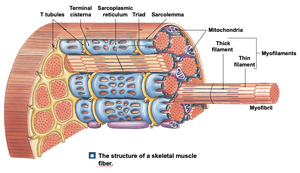
Structure of a Skeletal Muscle Fiber
Each skeletal muscle fiber is a long, multinucleated cell containing specialized structures for contraction and communication with nerves.
Sarcolemma: The plasma membrane of the muscle fiber.
Sarcoplasm: The cytoplasm of the muscle fiber, containing organelles and myofibrils.
Sarcoplasmic reticulum (SR): Modified endoplasmic reticulum that stores and releases calcium ions during contraction.
T-tubules (transverse tubules): Invaginations of the sarcolemma that transmit action potentials into the fiber.
Myofibrils: Bundles of actin and myosin filaments organized into sarcomeres.
Endomysium: Connective tissue surrounding each muscle fiber.
Key Terms and Structures
Important Terms
Sarcolemma: The cell membrane of a muscle fiber.
Sarcoplasm: The cytoplasm of a muscle fiber.
Sarcoplasmic reticulum: Stores calcium ions and releases them during muscle contraction.
Myofibrils: Cylindrical structures within the muscle fiber, composed of repeating sarcomeres.
Nuclei: Multiple, located at the periphery in skeletal muscle fibers.
T-tubules: Conduct action potentials from the sarcolemma to the interior of the muscle fiber.
Motor end plate: Specialized region of the sarcolemma at the neuromuscular junction, rich in acetylcholine receptors.
Neuromuscular junction (NMJ): The synapse between a motor neuron and a muscle fiber.
Neuromuscular Junction (NMJ)
Structure and Function
The neuromuscular junction is the site where a motor neuron communicates with a skeletal muscle fiber to initiate contraction. It consists of the axon terminal of the motor neuron, the synaptic cleft, and the motor end plate of the muscle fiber.
Synaptic cleft: The small gap between the neuron and muscle fiber.
Acetylcholine (ACh): The neurotransmitter released by the neuron to stimulate the muscle fiber.
Motor end plate: Contains receptors for ACh, initiating an action potential in the muscle fiber.
When an action potential reaches the axon terminal, ACh is released into the synaptic cleft, binds to receptors on the motor end plate, and triggers an influx of sodium ions. This depolarizes the sarcolemma, leading to muscle contraction.
Summary Table: Muscle Tissue Types
Muscle Type | Striations | Nuclei | Control | Location | Special Features |
|---|---|---|---|---|---|
Skeletal | Present | Multiple, peripheral | Voluntary | Attached to bones | Long, cylindrical fibers |
Cardiac | Present | Single, central | Involuntary | Heart | Branched fibers, intercalated discs |
Smooth | Absent | Single, central | Involuntary | Walls of hollow organs | Spindle-shaped cells |
Additional info: For more detailed study, refer to histology slides and models to identify the unique features of each muscle tissue type and the organization of skeletal muscle fibers. Understanding the NMJ is crucial for grasping how nerve impulses lead to muscle contraction.
