 Back
BackMuscle Tissue and Muscular System: Axial and Appendicular Muscles
Study Guide - Smart Notes
 Tailored notes based on your materials, expanded with key definitions, examples, and context.
Muscle Tissue
Types of Muscle Tissue
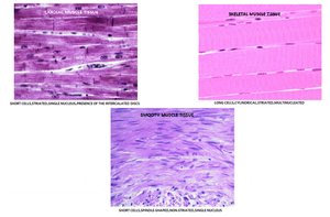
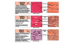
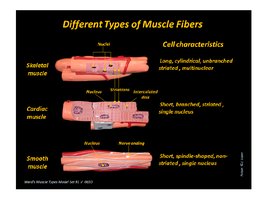
Muscle tissue is specialized for contraction and is essential for movement, posture, and vital functions. There are three main types of muscle tissue, each with distinct structural and functional characteristics:
Skeletal Muscle: Voluntary, striated, multinucleated, and attached to bones for movement.
Cardiac Muscle: Involuntary, striated, branched, and found only in the heart.
Smooth Muscle: Involuntary, non-striated, spindle-shaped, and found in walls of hollow organs.
Structure of Skeletal Muscle Cells
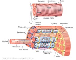
Skeletal muscle fibers are long, cylindrical cells containing multiple nuclei. Their internal structure is highly organized to facilitate contraction.
Myofibrils: Bundles of contractile proteins (actin and myosin).
Sarcolemma: The cell membrane of a muscle fiber.
Sarcoplasmic Reticulum: Specialized endoplasmic reticulum for calcium storage.
T Tubules: Invaginations of the sarcolemma that help transmit action potentials.
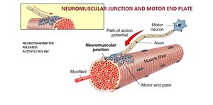
Neuromuscular Junction
The neuromuscular junction is the site where a motor neuron communicates with a muscle fiber, triggering contraction via the release of the neurotransmitter acetylcholine.
Motor End Plate: Specialized region of the muscle fiber membrane.
Action Potential: Electrical signal that initiates muscle contraction.
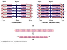
The Sarcomere: Functional Unit of Muscle Contraction
The sarcomere is the basic contractile unit of skeletal muscle, defined by Z lines. It contains thick (myosin) and thin (actin) filaments whose interaction produces contraction.
Sliding Filament Theory: Muscle contraction occurs as actin and myosin filaments slide past each other, shortening the sarcomere.
Bands and Zones: A band (thick filaments), I band (thin filaments), H zone (center of A band), Z line (boundary).
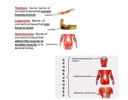
Tendons, Ligaments, and Aponeuroses
Connective Tissue Structures
Muscles are connected to bones and other structures via specialized connective tissues:
Tendons: Narrow bands connecting muscle to bone.
Ligaments: Bands connecting bone to bone.
Aponeuroses: Broad, flat sheets attaching muscles to other muscles or bones.
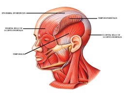
Axial Muscles
Scalp and Facial Muscles
Axial muscles include those of the head and neck, responsible for facial expression, mastication, and movement of the head.
Epicranial Aponeurosis: Connects frontal and occipital bellies of occipitofrontalis.
Temporalis: Elevates the mandible.
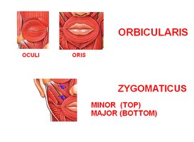
Facial Expression Muscles
Orbicularis Oculi: Closes the eyelids.
Orbicularis Oris: Closes and protrudes the lips.
Zygomaticus Major and Minor: Elevate the corners of the mouth.
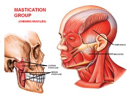
Muscles of Mastication
Masseter: Elevates the mandible.
Temporalis: Elevates and retracts the mandible.
Medial and Lateral Pterygoids: Move the jaw side-to-side.
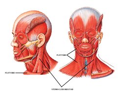
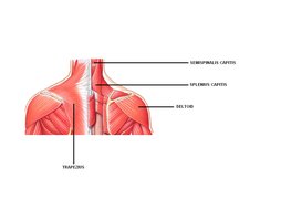
Neck Muscles
Sternocleidomastoid: Flexes and rotates the head.
Platysma: Tenses skin of the neck.
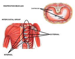
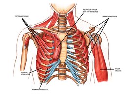
Muscles of Respiration
Diaphragm: Primary muscle of respiration.
Intercostal Muscles: External and internal groups assist with breathing.
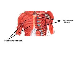
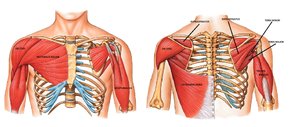
Muscles of the Trunk
Pectoralis Major and Minor: Move the shoulder and arm.
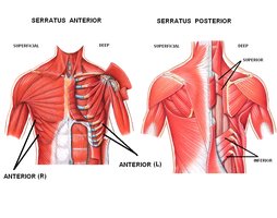
Serratus Anterior: Protracts the scapula.
Trapezius: Moves the scapula and supports the arm.
Latissimus Dorsi: Extends, adducts, and rotates the arm.
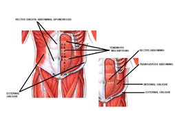
Abdominal Muscles and Aponeurosis
Rectus Abdominis: Flexes the vertebral column.
External and Internal Obliques: Rotate and flex the trunk.
Transversus Abdominis: Compresses abdominal contents.
Abdominal Aponeurosis: Broad connective tissue supporting abdominal muscles.
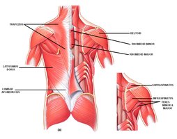
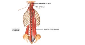
Vertebral Column Muscles
Erector Spinae: Extends and laterally flexes the vertebral column.
Quadratus Lumborum: Stabilizes the pelvis and lumbar spine.
Appendicular Muscles
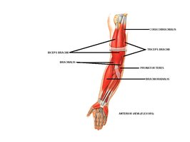
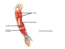
Upper Limb Muscles
Biceps Brachii: Flexes the elbow and supinates the forearm.
Triceps Brachii: Extends the elbow.
Brachialis: Flexes the elbow.
Brachioradialis: Flexes the forearm.
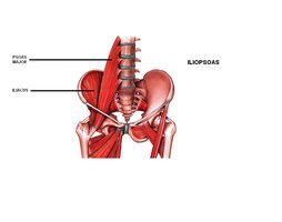
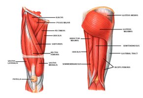
Lower Limb Muscles
Iliopsoas: Flexes the hip.
Gluteus Maximus and Medius: Extend and abduct the hip.
Quadriceps Group: Extends the knee.
Hamstring Group: Flexes the knee.
Gastrocnemius and Soleus: Plantar flex the foot.
Summary Table: Muscle Tissue Types
Type | Location | Control | Structure |
|---|---|---|---|
Skeletal | Attached to bones | Voluntary | Striated, multinucleated |
Cardiac | Heart | Involuntary | Striated, branched, single nucleus |
Smooth | Walls of organs | Involuntary | Non-striated, single nucleus |
Summary Table: Major Axial and Appendicular Muscles
Region | Muscle | Function |
|---|---|---|
Head/Face | Orbicularis oculi, orbicularis oris, zygomaticus, masseter, temporalis | Facial expression, mastication |
Neck | Sternocleidomastoid, platysma | Head movement, neck tension |
Trunk | Pectoralis major/minor, serratus anterior, trapezius, latissimus dorsi | Shoulder and arm movement |
Abdomen | Rectus abdominis, obliques, transversus abdominis | Trunk flexion, rotation, compression |
Back | Erector spinae, quadratus lumborum | Spine extension, stabilization |
Upper Limb | Biceps brachii, triceps brachii, brachialis, brachioradialis | Elbow flexion/extension |
Lower Limb | Iliopsoas, gluteus maximus/medius, quadriceps, hamstrings, gastrocnemius, soleus | Hip/knee movement, foot flexion |
Key Equations
Sliding Filament Theory:
Force Generation:
Additional info:
Muscle tissue is covered in detail in Ch. 10 (Muscle Tissue) and Ch. 11 (The Muscular System) of standard Anatomy & Physiology textbooks.
Axial muscles are those associated with the head, neck, and trunk; appendicular muscles are those of the limbs.
