 Back
Muscle Tissue and Organization: Study Notes for Anatomy & Physiology
Study Guide - Smart Notes
 Tailored notes based on your materials, expanded with key definitions, examples, and context.
Muscle Tissue Types
Overview of Muscle Tissue
Muscle tissue is essential for movement and the transport of materials within the body. There are three primary types of muscle tissue, each with distinct structural and functional characteristics.
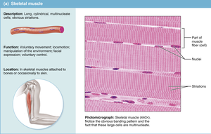
Skeletal Muscle: Striated, voluntary, multinucleate cells. Responsible for voluntary movements, locomotion, and manipulation of the environment. Found attached to bones or occasionally to skin.
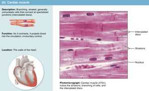
Cardiac Muscle: Striated, involuntary, usually uninucleate cells with intercalated discs. Propels blood through the heart and circulatory system. Located in the walls of the heart.
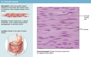
Smooth Muscle: Not striated, involuntary, spindle-shaped cells. Moves substances through hollow organs (e.g., digestive tract, blood vessels). Found mostly in the walls of hollow organs.
Properties of Muscle Tissue
Functional Characteristics
Muscle tissue exhibits four key properties that enable its function:
Excitability: Ability to respond to stimuli, typically from the nervous system.
Contractility: Ability to shorten and generate force, resulting in movement or tension.
Elasticity: Ability to return to original length after contraction or extension.
Extensibility: Ability to stretch beyond resting length in response to opposing muscle contraction.
Functions of Skeletal Muscle
Major Roles in the Body
Skeletal muscle performs several vital functions:
Body Movement: Muscles pull across joints to move the body.
Posture and Joint Stabilization: Continuous contraction maintains posture and stabilizes joints.
Heat Production: Muscle activity generates heat, helping regulate body temperature.
Control of Passageways: Sphincter muscles open and close body passageways.
Muscle Structural Organization
Hierarchical Structure
Skeletal muscle is organized in a hierarchical manner:
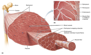
Whole Muscle → Muscle Fascicles → Muscle Fibers → Myofibrils → Myofilaments (actin/myosin, arranged in sarcomeres)
Connective Tissues of Skeletal Muscle
Three Layers of Connective Tissue
Connective tissue layers provide structural support and attachment to the skeleton:
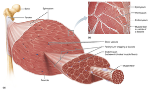
Epimysium: Dense irregular connective tissue surrounding the whole muscle.
Perimysium: Dense irregular connective tissue sheath around fascicles; contains blood vessels and nerves.
Endomysium: Areolar connective tissue around each muscle fiber.
Nerves and Blood Vessels
Innervation and Vascular Supply
Each skeletal muscle receives one nerve, one artery, and one or more veins, which branch extensively within the muscle. Capillaries in the endomysium form a network to meet the high demand for nutrients and oxygen.
Muscle Attachments
Tendons and Aponeuroses
The connective tissue layers merge to form tendons (thick cords) or aponeuroses (flat sheets) for muscle attachment to bones.
Origin: Less movable, usually proximal attachment.
Insertion: More movable, usually distal attachment; moves toward origin during contraction.
Composition of Tendons
Structure and Function
Tendons are composed of tenocytes, water, and fibrous collagen proteins. Collagen fibers form strong, flexible strands that connect muscle to bone, creating a durable and permanent bond.
Skeletal Muscle Fiber Structure
Myofibrils and Sarcomeres
Skeletal muscle fibers are multinucleate and contain myofibrils, which are composed of repeating units called sarcomeres. Sarcomeres are the functional contractile units, giving muscle its striated appearance.
Sarcomere Structure and Function
Microscopic Anatomy
Z discs: Protein boundaries of sarcomeres.
Actin: Thin filaments extending from Z discs toward the center.
Myosin: Thick filaments connected to Z discs by titin.
Sarcoplasmic Reticulum and T Tubules
Calcium Regulation and Muscle Contraction
The sarcoplasmic reticulum (SR) is a smooth endoplasmic reticulum that surrounds the muscle fiber. T tubules are deep invaginations of the sarcolemma, flanked by terminal cisterns of the SR. Muscle contraction is triggered when a motor neuron signal causes the release of calcium from the SR, activating the sarcomere.
Skeletal Muscle Contraction
Types of Contraction
Concentric Contraction: Muscle shortens while generating force (sliding filament mechanism).
Eccentric Contraction: Muscle lengthens while generating force (controlled movements, resistance to gravity).
Muscle Extension: Stretching reduces overlap of thick and thin filaments.
Motor Units and Innervation
Neural Control of Muscle
Skeletal muscles are voluntary, controlled by the somatic nervous system. Motor neurons innervate muscle fibers, forming motor units. The axon transmits impulses through connective tissue layers to individual muscle fibers.
Types of Skeletal Muscle Fibers
Classification by ATP Production and Contraction Speed
Slow Oxidative (SO): Aerobic ATP production, high myoglobin, slow contraction, postural muscles.
Fast Oxidative (FO): Intermediate, fast contraction, aerobic ATP production, some myoglobin.
Fast Glycolytic (FG): Anaerobic ATP production, low myoglobin, fast contraction, large diameter, prevalent in muscles requiring quick, powerful movements.
Fiber Distribution and Genetics
Variation Among Muscles and Individuals
The proportion of fiber types varies by muscle and individual genetics. SO fibers dominate postural muscles; FG fibers are more common in sprinters. Training can alter fiber function, but genetic proportions remain constant.
Disorders of Skeletal Muscle
Common Muscle Disorders
Muscular Dystrophy: Inherited disease causing muscle degeneration and replacement by fat/connective tissue.
Duchenne Muscular Dystrophy: Sex-linked, early onset, progressive degeneration.
Myotonic Dystrophy: Spasms, weakness, abnormal heart rhythm.
Myofascial Pain: Pain from tight muscle bands, often in postural muscles.
Fibromyalgia: Chronic pain of unknown cause, common in women, diagnosed by standardized points.
Aging in Skeletal Muscle
Effects of Aging
Progressive loss of muscle mass begins in mid-30s due to inactivity.
Lost muscle replaced by adipose or fibrous tissue.
Decreased size, power, strength, and endurance.
Reduced cardiovascular performance and slower circulatory supply.
Lower tolerance for exercise and rapid fatigue.
Reduced recovery capacity and elasticity.
