 Back
BackMuscle Tissue and Physiology: Structure and Function of the Muscular System
Study Guide - Smart Notes
 Tailored notes based on your materials, expanded with key definitions, examples, and context.
The Muscular System
Overview of Skeletal Muscle Structure
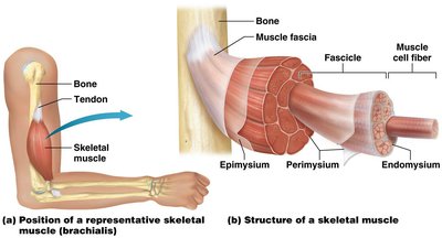
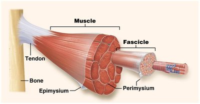
The muscular system is composed of skeletal muscles, which are responsible for voluntary movements, posture, and heat production. Each skeletal muscle is a complex organ containing muscle fibers, connective tissues, blood vessels, and nerves.
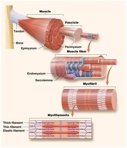
Muscle fibers (cells) are bundled into fascicles, which are grouped together to form the whole muscle.
Connective tissue layers include the endomysium (surrounds individual muscle fibers), perimysium (surrounds fascicles), and epimysium (surrounds the entire muscle).
Muscles attach to bones via tendons, allowing movement of the skeleton.
Functional Groups of Muscles
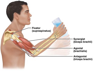
Muscles rarely act alone; instead, they work in groups to produce coordinated movements. Each group has specific roles:
Agonist (prime mover): Main muscle responsible for a movement.
Antagonist: Opposes the action of the agonist, allowing for controlled movement.
Synergist: Assists the agonist by adding force or reducing unwanted movement.
Fixator: Stabilizes the origin of the agonist to increase efficiency and prevent injury.
Muscle Origin and Insertion
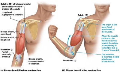
Each skeletal muscle has a fixed point of attachment called the origin and a movable point called the insertion. During contraction, the insertion moves toward the origin.
Origin: Typically proximal and less movable.
Insertion: Typically distal and more movable.
Muscle Tissue Types
Overview of Muscle Tissue
There are three types of muscle tissue in the human body, each with distinct structural and functional characteristics:
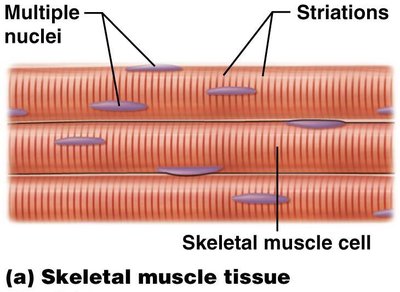
Skeletal muscle: Voluntary, striated, multinucleated, attached to bones.
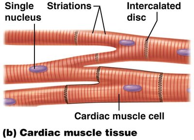
Cardiac muscle: Involuntary, striated, single nucleus, found in the heart, contains intercalated discs.
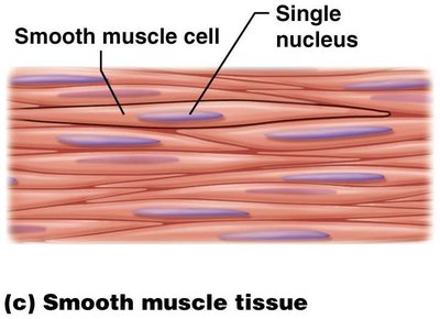
Smooth muscle: Involuntary, non-striated, single nucleus, found in walls of hollow organs.
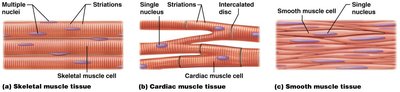
Skeletal Muscle Tissue
Shape: Long, cylindrical fibers
Striations: Present
Number of nuclei: Multiple, peripherally located
Control: Voluntary
Location: Attached to bones
Cardiac Muscle Tissue
Shape: Branched fibers
Striations: Present
Number of nuclei: Single, centrally located
Control: Involuntary
Location: Heart
Special feature: Intercalated discs for synchronized contraction
Smooth Muscle Tissue
Shape: Spindle-shaped fibers
Striations: Absent
Number of nuclei: Single, centrally located
Control: Involuntary
Location: Walls of hollow organs (e.g., intestines, blood vessels)
Properties and Structure of Muscle Cells
Properties of Muscle Cells
Muscle cells (myocytes) possess unique properties that enable their function:
Contractility: Ability to shorten and generate force.
Excitability: Ability to respond to stimuli.
Conductivity: Ability to conduct electrical signals.
Extensibility: Ability to stretch without damage.
Elasticity: Ability to return to original length after stretching.
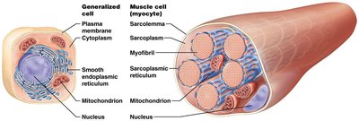
Structure of Muscle Cells (Myocytes)
Muscle cells are highly specialized for contraction and contain several unique structures:
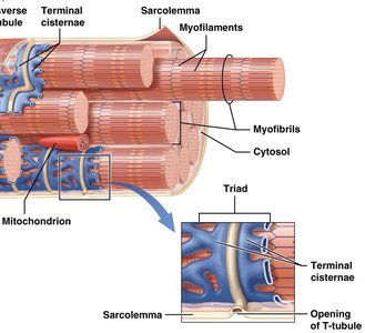
Sarcoplasm: Cytoplasm of the muscle cell.
Sarcolemma: Plasma membrane of the muscle cell.
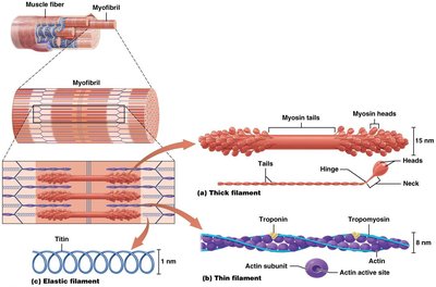
Myofibrils: Bundles of contractile proteins (actin and myosin).
Sarcoplasmic reticulum (SR): Modified smooth endoplasmic reticulum that stores calcium ions.
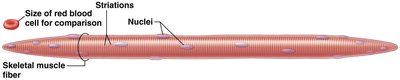
Structure of the Skeletal Muscle Fiber
Skeletal muscle fibers are long, cylindrical cells with multiple nuclei and visible striations. They are much larger than most other cells in the body.
Transverse Tubules and Myofibril Structure
Transverse tubules (T-tubules) are invaginations of the sarcolemma that help transmit action potentials deep into the muscle fiber. Myofibrils are composed of three types of myofilaments:
Thick filaments: Composed of myosin.
Thin filaments: Composed of actin, tropomyosin, and troponin.
Elastic filaments: Composed of titin, providing elasticity and stability.
Duchenne Muscular Dystrophy (DMD)
Pathophysiology of DMD
Duchenne muscular dystrophy is a genetic disorder caused by a defective gene for the protein dystrophin. Dystrophin anchors the sarcolemma to the surrounding connective tissue and myofibrils. Without functional dystrophin, muscle fibers degenerate and are replaced by fat and connective tissue.
Symptoms: Muscle weakness, especially in proximal limb muscles, waddling gait, loss of ambulation by age 12, and early death due to respiratory or cardiac failure.
The Big Picture: Skeletal Muscle Structure
Hierarchical Organization
Skeletal muscle is organized in a hierarchical manner:
Muscle fibers are grouped into fascicles, surrounded by perimysium.
Fascicles are bundled together to form the muscle, surrounded by epimysium.
The connective tissue layers converge to form tendons, which attach muscle to bone.
Fascia surrounds and separates groups of muscles.
Levels of Muscle Structure
Muscle → Fascicle → Muscle fiber → Myofibril → Myofilament
Sarcomere Structure and Muscle Contraction
Sarcomere Organization
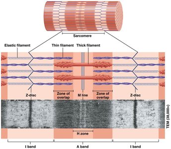
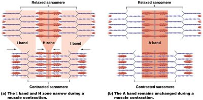
The sarcomere is the functional unit of muscle contraction, defined by the region between two Z-discs. It contains overlapping thick and thin filaments, creating striations.
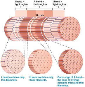
I band: Contains only thin filaments.
A band: Contains both thick and thin filaments.
H zone: Contains only thick filaments.
Z disc: Defines the boundary of each sarcomere.
M line: Center of the sarcomere.
Sliding Filament Theory
The sliding filament theory explains how muscles contract:
During contraction, thin filaments slide past thick filaments, shortening the sarcomere.
The I band and H zone narrow, while the A band remains unchanged.
Z-discs move closer together, shortening the muscle fiber.
Neuromuscular Junction and Muscle Contraction
Neuromuscular Junction (NMJ)
The NMJ is the synapse between a motor neuron and a skeletal muscle fiber. It is essential for initiating muscle contraction.
Motor neuron: Sends the signal to contract.
Axon terminal: Releases acetylcholine (ACh) into the synaptic cleft.
Motor end plate: Specialized region of the muscle membrane with ACh receptors.
Phases of Muscle Contraction
Excitation: Action potential triggers ACh release, leading to depolarization of the muscle fiber.
Excitation-contraction coupling: Muscle action potential triggers Ca2+ release from the SR, exposing actin active sites.
Contraction: Myosin heads bind to actin, perform power strokes, and slide filaments past each other using ATP.
Summary Table: Muscle Tissue Types
Muscle Type | Striations | Nuclei | Control | Location | Special Features |
|---|---|---|---|---|---|
Skeletal | Yes | Multiple, peripheral | Voluntary | Attached to bones | Long fibers |
Cardiac | Yes | Single, central | Involuntary | Heart | Intercalated discs |
Smooth | No | Single, central | Involuntary | Walls of hollow organs | Spindle-shaped |
