 Back
BackMuscle Tissue and Physiology: Structure and Function of the Muscular System
Study Guide - Smart Notes
 Tailored notes based on your materials, expanded with key definitions, examples, and context.
Muscle Tissue and Physiology
Introduction to Muscle Tissue
Muscle tissue is essential for movement, posture, and various physiological processes. There are three main types of muscle tissue: skeletal, cardiac, and smooth. Each type has unique structural and functional characteristics that enable the body to perform a wide range of activities.
The Muscular System
Overview of the Muscular System
The muscular system consists of all the skeletal muscles in the body, which are responsible for voluntary movements, maintaining posture, and supporting soft tissues. Muscles work in groups to produce coordinated actions and are attached to bones via tendons.
Structure of Skeletal Muscle
Organization of Skeletal Muscle
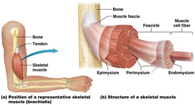
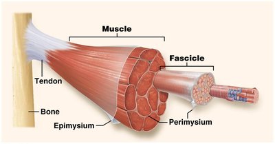
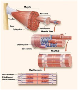
Skeletal muscle is composed of muscle fibers (cells), connective tissue, blood vessels, and nerves. The connective tissue layers organize muscle fibers into bundles and connect muscles to bones.
Endomysium: Surrounds each individual muscle fiber.
Perimysium: Bundles several muscle fibers into a fascicle.
Epimysium: Encloses all fascicles to form the complete muscle.
Tendons: Connect muscle to bone, formed by the merging of connective tissue layers.
Functional Groups of Muscles
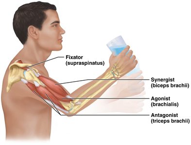
Muscles rarely act alone; they function in groups to produce movement and maintain stability.
Agonist (Prime Mover): Provides the main force for a movement.
Antagonist: Opposes the action of the agonist, allowing for controlled movement.
Synergist: Assists the agonist by adding force or reducing unwanted movement.
Fixator: Stabilizes the origin of the agonist to prevent unnecessary movement.
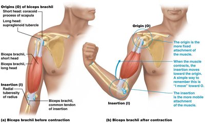
Muscle Origin and Insertion
Each skeletal muscle has a specific origin and insertion point:
Origin: The fixed attachment point, usually on a bone, that does not move during contraction.
Insertion: The movable attachment point, where the muscle exerts its action.
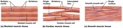
Types of Muscle Tissue
Comparison of Muscle Tissue Types
There are three types of muscle tissue, each with distinct structural and functional properties:
Type | Shape | Striations | Nuclei | Control | Location |
|---|---|---|---|---|---|
Skeletal | Long, cylindrical | Present | Multiple | Voluntary | Attached to bones |
Cardiac | Branched | Present | Single | Involuntary | Heart |
Smooth | Spindle-shaped | Absent | Single | Involuntary | Walls of hollow organs |
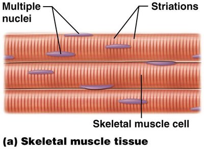
Skeletal Muscle Tissue
Shape: Long, cylindrical fibers
Striations: Present
Number of Nuclei: Multiple, peripherally located
Control: Voluntary
Location: Attached to bones
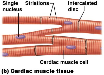
Cardiac Muscle Tissue
Shape: Branched fibers
Striations: Present
Number of Nuclei: Single, centrally located
Control: Involuntary
Location: Heart
Special Feature: Intercalated discs for cell-to-cell communication
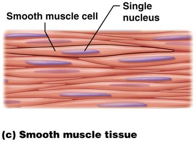
Smooth Muscle Tissue
Shape: Spindle-shaped fibers
Striations: Absent
Number of Nuclei: Single, centrally located
Control: Involuntary
Location: Walls of hollow organs (e.g., intestines, blood vessels)
Properties and Structure of Muscle Cells
Properties of Muscle Cells
Contractility: Ability to shorten and generate force.
Excitability: Ability to respond to stimuli.
Conductivity: Ability to conduct electrical signals.
Extensibility: Ability to stretch without damage.
Elasticity: Ability to return to original length after stretching.
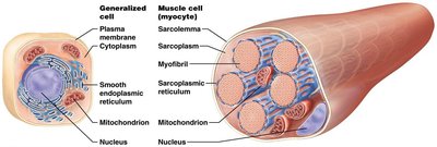
Structure of Muscle Cells (Myocytes)
Sarcoplasm: Cytoplasm of the muscle cell.
Sarcolemma: Plasma membrane of the muscle cell.
Myofibrils: Bundles of contractile proteins within the cell.
Sarcoplasmic Reticulum (SR): Modified smooth endoplasmic reticulum that stores calcium ions.
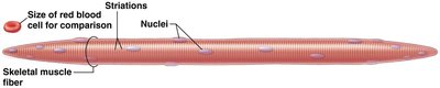
Structure of the Skeletal Muscle Fiber
Skeletal muscle fibers are long, cylindrical cells with multiple nuclei and visible striations. They are much larger than most other cells in the body.
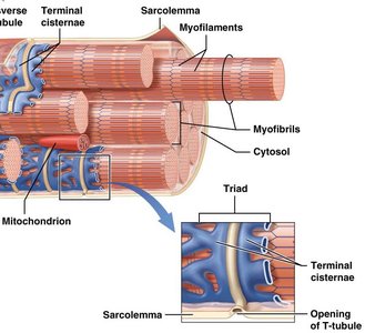
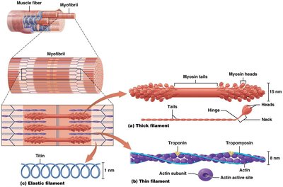
Transverse Tubules and Myofibril Structure
Transverse tubules (T-tubules) are invaginations of the sarcolemma that help transmit action potentials deep into the muscle fiber. Myofibrils are composed of three types of myofilaments:
Thick filaments: Made of myosin
Thin filaments: Made of actin, tropomyosin, and troponin
Elastic filaments: Made of titin
Duchenne Muscular Dystrophy (DMD)
Overview of DMD
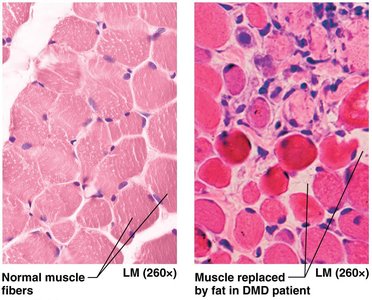
Duchenne Muscular Dystrophy is a genetic disorder caused by a defective gene for the protein dystrophin. This protein anchors the sarcolemma to the surrounding connective tissue and myofibrils. Without functional dystrophin, muscle fibers degenerate and are replaced by fat and connective tissue.
Symptoms: Muscle weakness, especially in proximal limb muscles, waddling gait, loss of ambulation by age 12, and early death due to respiratory or cardiac failure.
The Big Picture of Skeletal Muscle Structure
Hierarchical Organization
Skeletal muscle is organized in a hierarchical manner:
Muscle fibers are surrounded by endomysium and grouped into fascicles (perimysium).
Fascicles are bundled together and surrounded by epimysium to form the whole muscle.
The connective tissue layers merge to form tendons, which attach muscle to bone.
Fascia surrounds groups of muscles, anchoring them to surrounding tissues.
Sarcomere Structure and Function
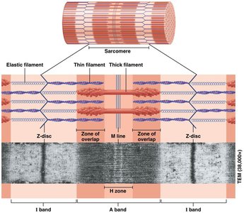
Sarcomere Organization
The sarcomere is the functional unit of muscle contraction, defined by the region between two Z-discs. It contains overlapping thick and thin filaments, which create the striated appearance of skeletal and cardiac muscle.
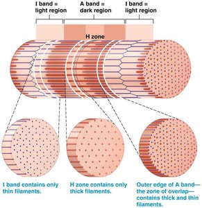
I band: Contains only thin filaments
A band: Contains both thick and thin filaments
H zone: Contains only thick filaments
Z disc: Defines the boundary of each sarcomere
M line: Center of the sarcomere
The Sliding Filament Theory
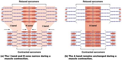
Mechanism of Muscle Contraction
The sliding filament theory explains how muscles contract by the sliding of thin filaments past thick filaments, shortening the sarcomere without changing the length of the filaments themselves.
The I band and H zone narrow during contraction.
The A band remains unchanged.
Z-discs move closer together, shortening the muscle fiber.
The Neuromuscular Junction (NMJ) and Muscle Contraction
Structure and Function of the NMJ
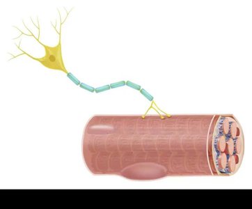
The neuromuscular junction is the synapse between a motor neuron and a skeletal muscle fiber. It is the site where nerve impulses trigger muscle contraction.
Motor neuron: Sends the signal to contract.
Synaptic vesicles: Contain acetylcholine (ACh), the neurotransmitter.
Synaptic cleft: Space between neuron and muscle fiber.
Motor end plate: Specialized region of the muscle membrane with ACh receptors.
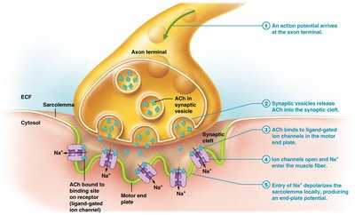
Phases of Muscle Contraction
Excitation: Action potential triggers ACh release, which binds to receptors and generates a muscle action potential.
Excitation-Contraction Coupling: Muscle action potential causes Ca2+ release from the SR, exposing active sites on actin.
Contraction: Myosin heads bind to actin, perform a power stroke, and cycle as long as ATP and Ca2+ are available.
Muscle Relaxation
Mechanisms of Relaxation
Acetylcholinesterase (AChE) degrades ACh in the synaptic cleft.
ATP breaks crossbridges between actin and myosin.
Calcium ions are pumped back into the SR.
Troponin and tropomyosin block actin active sites, ending contraction.
Rigor Mortis
Postmortem Muscle Contraction
Rigor mortis is the stiffening of muscles after death due to the lack of ATP, which prevents the detachment of myosin heads from actin. This state persists until muscle proteins degrade.
Sources of Energy for Muscle Contraction
ATP Generation Pathways
Immediate: Stored ATP and creatine phosphate (lasts ~10 seconds).
Glycolytic (Anaerobic): Glycolysis in the cytosol, produces ATP for 30–40 seconds.
Oxidative (Aerobic): Occurs in mitochondria, produces ATP for prolonged activity.
Muscle Fiber Types
Classification of Skeletal Muscle Fibers
Type | Diameter | Speed | Metabolism | Fatigue Resistance | Color |
|---|---|---|---|---|---|
Type I | Small | Slow | Oxidative | High | Red |
Type IIa | Intermediate | Fast | Oxidative-Glycolytic | Intermediate | Pale |
Type IIx/IIb | Large | Fastest | Glycolytic | Low | White |
Motor Units and Muscle Contraction Types
Motor Units
A motor unit consists of a single motor neuron and all the muscle fibers it innervates. The size of the motor unit determines the precision of movement.
Small motor units: Fine control (e.g., fingers)
Large motor units: Gross movements (e.g., legs)
Types of Muscle Contractions
Isotonic Concentric: Muscle shortens while generating tension.
Isotonic Eccentric: Muscle lengthens while maintaining tension.
Isometric: Muscle length remains unchanged while generating tension.
Adaptations and Disorders of Muscle Tissue
Physical Training Effects
Endurance Training: Increases oxidative capacity, mitochondria, and blood supply.
Resistance Training: Increases muscle fiber size (hypertrophy) and myofibril number.
Disuse: Leads to atrophy, decreased strength, and endurance.
Muscle Fatigue
Fatigue is the inability to maintain a given level of intensity due to depletion of energy sources, decreased oxygen, or environmental factors.
Smooth and Cardiac Muscle
Smooth Muscle
Found in walls of hollow organs
No striations; actin and myosin arranged differently
Single-unit and multi-unit types
Cardiac Muscle
Found only in the heart
Branched, striated cells with intercalated discs
Autorhythmic due to pacemaker cells
