 Back
BackMuscle Tissue and Physiology: Structure, Function, and Contraction
Study Guide - Smart Notes
 Tailored notes based on your materials, expanded with key definitions, examples, and context.
Muscle Tissue
Overview of Muscle Tissue
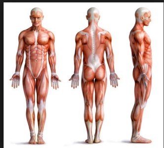
Muscle tissue constitutes nearly half of the body's mass and is essential for transforming chemical energy (ATP) into mechanical energy, enabling force generation and movement. There are three primary types of muscle tissue: skeletal, cardiac, and smooth muscle. Prefixes such as myo, mys, and sarco are commonly used in muscle terminology.
Skeletal muscle: Attached to bones and skin, composed of elongated, striated muscle fibers, under voluntary control, contracts rapidly and powerfully, but tires easily.
Cardiac muscle: Found only in the heart, striated, involuntary, can contract without nervous system stimulation.
Smooth muscle: Located in walls of hollow organs (e.g., stomach, bladder, airways), not striated, involuntary, can contract without nervous system stimulation.
Special Characteristics of Muscle Tissue
Muscle tissue exhibits four key properties:
Excitability (responsiveness): Ability to receive and respond to stimuli.
Contractility: Ability to shorten forcibly when stimulated.
Extensibility: Ability to be stretched.
Elasticity: Ability to recoil to resting length after stretching.
Functions of Muscle Tissue
Muscle tissue performs several vital functions:
Movement: Moves bones or fluids (e.g., blood).
Posture: Maintains posture and body position.
Joint stabilization: Stabilizes joints.
Heat generation: Generates heat, especially skeletal muscle.
Additional functions: Protects organs, forms valves, controls pupil size, causes "goosebumps".
Skeletal Muscle Structure
Connective Tissue Sheaths
Skeletal muscle is organized and supported by three layers of connective tissue:
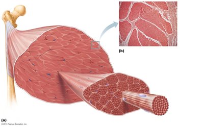
Epimysium: Dense irregular connective tissue surrounding the entire muscle; may blend with fascia.
Perimysium: Fibrous connective tissue surrounding fascicles (groups of muscle fibers).
Endomysium: Fine areolar connective tissue surrounding each muscle fiber.
Microscopic Anatomy of Skeletal Muscle Fiber
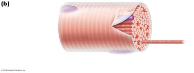
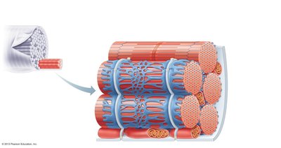
Skeletal muscle fibers are long, cylindrical cells with multiple peripheral nuclei. The sarcolemma is the plasma membrane, and the sarcoplasm is the cytoplasm, containing glycosomes (for glycogen storage) and myoglobin (for O2 storage). Modified structures include myofibrils, sarcoplasmic reticulum, and T tubules.
Myofibrils and Sarcomeres
Myofibrils
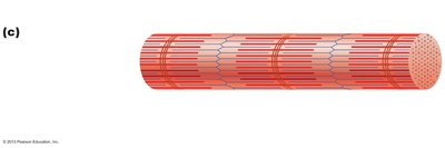
Myofibrils are densely packed, rod-like elements that make up about 80% of the cell volume. They contain sarcomeres, the functional contractile units of skeletal muscle cells, which are composed of myofilaments. Myofibrils exhibit striations due to the perfectly aligned repeating series of dark A bands and light I bands.
Structure and Organizational Levels of Skeletal Muscle
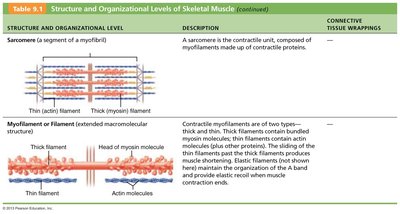
The sarcomere is the contractile unit, composed of myofilaments made up of contractile proteins. Myofilaments are classified as thick (myosin) and thin (actin) filaments. The sliding of thin filaments past thick filaments produces muscle shortening.
Structure and Organizational Level | Description |
|---|---|
Sarcomere | Segment of a myofibril; contractile unit composed of myofilaments |
Myofilament | Thick (myosin) and thin (actin) filaments; responsible for contraction |
Banding Pattern and Sarcomere Structure
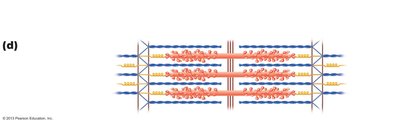
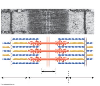
The sarcomere is the smallest contractile unit of muscle fiber, aligned along myofibrils like boxcars of a train. It contains an A band with half an I band at each end and is composed of thick and thin myofilaments made of contractile proteins.
H zone: Lighter region in midsection of dark A band where filaments do not overlap.
M line: Line of protein myomesin bisects H zone.
Z disc: Coin-shaped sheet of proteins on midline of light I band that anchors thin filaments and connects myofibrils.
Thick filaments: Run entire length of A band.
Thin filaments: Run length of I band and partway into A band.
Ultrastructure of Myofilaments
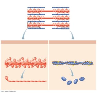
Thick filament: Composed of myosin protein (tails and heads). Myosin heads act as cross bridges during contraction, with binding sites for actin and ATP.
Thin filament: Twisted double strands of actin protein, bearing active sites for myosin head attachment. Regulatory proteins tropomyosin and troponin are bound to actin.
Elastic filament: Composed of titin protein, holds thick filaments in place, helps recoil after stretch, resists excessive stretching.
Sarcoplasmic Reticulum and T Tubules
Sarcoplasmic Reticulum (SR)
The SR is a network of smooth endoplasmic reticulum surrounding each myofibril, running mostly longitudinally. Pairs of terminal cisterns form perpendicular cross channels. The SR regulates intracellular Ca2+ levels, storing and releasing Ca2+ as needed for muscle contraction.
T Tubules
T tubules are continuations of the sarcolemma, with their lumen continuous with extracellular space. They penetrate the cell's interior at each A band–I band junction and associate with paired terminal cisterns to form triads that encircle each sarcomere.
Triad Relationships
T tubules conduct impulses deep into muscle fiber, reaching every sarcomere.
Integral proteins protrude into intermembrane space from T tubule and SR cistern membranes, connecting with each other.
T tubule integral proteins act as voltage sensors, changing shape in response to voltage changes.
SR integral proteins are channels that release Ca2+ from SR cisterns when voltage sensors change shape.
Sliding Filament Model of Contraction
Mechanism of Contraction
Muscle contraction is based on the sliding filament model:
Force is generated when tension from cross bridges on thin filaments exceeds forces opposing shortening.
In relaxed state, thin and thick filaments overlap only at ends of A band.
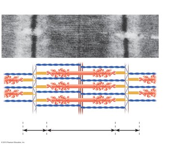
During contraction, thin filaments slide past thick filaments, increasing actin and myosin overlap.
Myosin heads bind to actin, forming cross bridges, which ratchet thin filaments toward the center of the sarcomere.
This causes shortening of the muscle fiber, pulling Z discs toward the M line, shortening I bands, bringing Z discs closer, causing H zones to disappear, and A bands to move closer (length stays the same).
Physiology of Skeletal Muscle Fibers
Requirements for Contraction
Activation (at neuromuscular junction): Requires nervous system stimulation and generation of action potential in sarcolemma (end-plate potential).
Excitation-contraction coupling: Action potential propagates along sarcolemma, intracellular Ca2+ levels rise briefly, cross bridge formation occurs between actin and myosin heads.
Phases Leading to Muscle Fiber Contraction
Action potential arrives at axon terminal at neuromuscular junction.
ACh released; binds to receptors on sarcolemma.
Ion permeability of sarcolemma changes.
Local change in membrane voltage (depolarization) occurs.
Local depolarization (end plate potential) ignites action potential in sarcolemma.
Action potential travels across entire sarcolemma and along T tubules.
SR releases Ca2+; Ca2+ binds to troponin; myosin-binding sites on actin exposed.
Myosin heads bind to actin; contraction begins.
Neuromuscular Junction (NMJ)
Structure and Function
Skeletal muscles are stimulated by somatic motor neurons.
Axons of motor neurons travel from the CNS via nerves to skeletal muscle, forming several branches (axon terminals) as they enter muscle.
Each axon ending forms a neuromuscular junction with a single muscle fiber, usually only one per muscle fiber.
NMJ is situated midway along the length of the muscle fiber.
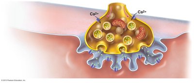
Axon terminal and sarcolemma are separated by a gel-filled space called the synaptic cleft.
Synaptic vesicles contain acetylcholine (ACh), a neurotransmitter.
Junctional folds of sarcolemma contain ACh-specific (ligand-gated) receptors.
NMJ includes axon terminals, synaptic cleft, and junctional folds of sarcolemma.
Events at the Neuromuscular Junction
Action potential arrives at axon terminal of motor neuron.
Voltage-gated Ca2+ channels open; Ca2+ enters the axon terminal.
Ca2+ entry causes ACh to be released by exocytosis.
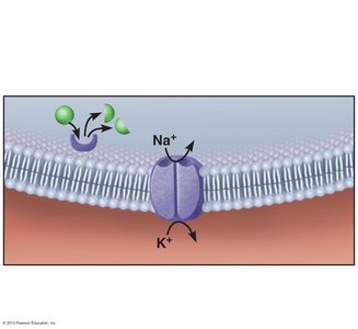
ACh diffuses across the synaptic cleft and binds to its receptors on the sarcolemma.
ACh binding opens ion channels in the receptors, allowing simultaneous passage of Na+ into the muscle fiber and K+ out. More Na+ enters than K+ exits, producing a local change in membrane potential called the end plate potential.
ACh effects are terminated by its breakdown in the synaptic cleft by acetylcholinesterase and diffusion away from the junction.
Summary of Events at the Neuromuscular Junction
Nerve impulse arrives at axon terminal, causing ACh release into synaptic cleft.
ACh diffuses across cleft and binds with receptors on sarcolemma.
Electrical events generate a local action potential called the end plate potential.
Additional info: The notes above expand on the original content by providing definitions, examples, and context for each structure and process, ensuring a comprehensive and self-contained study guide for exam preparation in anatomy and physiology.
