 Back
BackMuscle Tissue and Physiology: Structure, Function, and Mechanisms
Study Guide - Smart Notes
 Tailored notes based on your materials, expanded with key definitions, examples, and context.
Muscle Tissue and Physiology
Overview of Muscle Tissue
Muscle tissue is essential for movement, posture, joint stability, heat generation, and regulation of material flow through hollow organs. It consists of specialized cells called myocytes and a surrounding extracellular matrix known as the endomysium. There are three main types of muscle tissue: skeletal, cardiac, and smooth.
Muscle Tension: The force generated by muscle tissue, responsible for movement and other physiological functions.
Myocytes: Muscle cells that contract to produce force.
Endomysium: Connective tissue that holds muscle cells together and transmits tension.
Types of Muscle Tissue
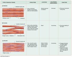
Muscle tissue is classified based on structure, location, control, and function.
Skeletal Muscle: Long, multinucleated, striated fibers; voluntary control; attached to skeleton; responsible for body movement.
Cardiac Muscle: Short, branched, striated cells with one nucleus; involuntary; found only in the heart; contracts as a unit due to intercalated discs.
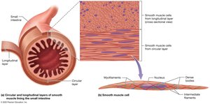
Smooth Muscle: Spindle-shaped, non-striated cells with one nucleus; involuntary; found in hollow organs, eyes, skin, and ducts; controls flow and movement within organs.
Properties of Muscle Cells
Muscle cells possess unique physiological properties that enable their function:
Contractility: Ability to shorten and generate force.
Excitability: Ability to respond to stimuli (chemical, mechanical, or electrical).
Conductivity: Ability to conduct electrical signals across the membrane.
Distensibility: Ability to stretch without rupture.
Elasticity: Ability to return to original length after stretching.
Structure of Muscle Cells
General Structure
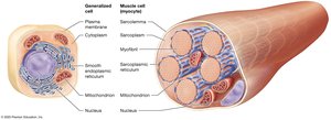
Muscle cells share many organelles with other cells but have specialized structures for contraction:
Sarcoplasm: Cytoplasm of a muscle cell.
Sarcolemma: Plasma membrane of a muscle cell.
Myofibrils: Bundles of contractile proteins, making up most of the cell's volume.
Sarcoplasmic Reticulum (SR): Modified smooth endoplasmic reticulum that stores and releases calcium ions.
Skeletal Muscle Fiber Structure
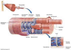
Skeletal muscle fibers are long, cylindrical, multinucleated, and striated. They arise from the fusion of embryonic myoblasts.
Size: About 100 µm in diameter and up to 30 cm long.
Striations: Alternating light and dark bands visible under a microscope.
Multiple Nuclei: Result from myoblast fusion.

Internal Organization
Key features of skeletal muscle fibers include:
Myofibrils: Surrounded by the sarcoplasmic reticulum, which stores and releases calcium.
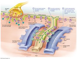
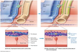
Transverse Tubules (T-tubules): Inward extensions of the sarcolemma that surround each myofibril and are filled with extracellular fluid.
Terminal Cisternae: Enlarged regions of the SR flanking each T-tubule; together with a T-tubule, they form a triad.
Myofibril and Myofilament Structure
Types of Myofilaments
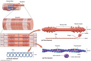
Myofibrils are composed of three types of myofilaments:
Thick Filaments: Made of myosin, with globular heads and a tail; responsible for force generation.
Thin Filaments: Composed of actin (with active sites), tropomyosin (covers active sites at rest), and troponin (regulates contraction).
Elastic Filaments: Made of titin, providing elasticity and structural support.
Sarcomere Organization
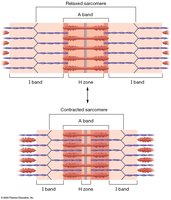
The sarcomere is the functional unit of muscle contraction, defined by the arrangement of myofilaments:
I Band: Light region; contains only thin filaments.
A Band: Dark region; contains thick filaments (and overlapping thin filaments).
H Zone: Central region of A band; only thick filaments.
M Line: Middle of A band; holds thick filaments in place.
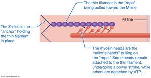
Z-Disc: Anchors thin filaments and marks the boundary of each sarcomere.
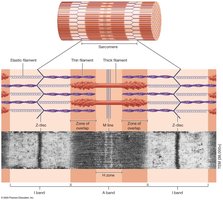
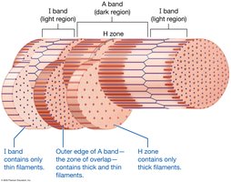
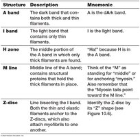
Structure | Description | Mnemonic |
|---|---|---|
A band | Dark band with thick and thin filaments | A is the dArk band |
I band | Light band with only thin filaments | I is the lIght band |
H zone | Middle of A band, only thick filaments | "H" is in the A band |
M line | Middle of A band, holds thick filaments | M for "middle" or "myosin" |
Z-disc | Bisects I band, anchors filaments | Z shape (see figure) |
Levels of Organization
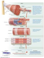
Muscle structure is organized hierarchically from the whole muscle down to myofilaments:
Muscle → Fascicle → Muscle Fiber → Myofibril → Myofilament
Sliding-Filament Mechanism of Contraction
Mechanism
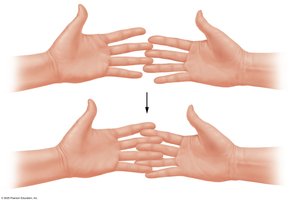
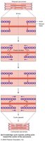
Muscle contraction occurs as thin filaments slide past thick filaments, shortening the sarcomere and generating tension. The I bands and H zone narrow, while the A band remains unchanged.
Membrane Potential and Action Potentials
Resting Membrane Potential
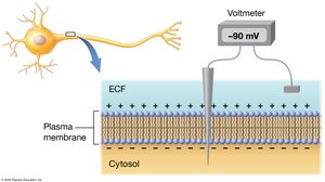
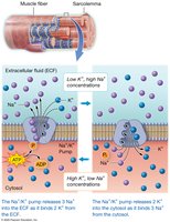
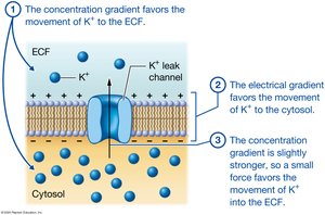
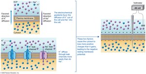
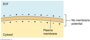
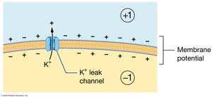
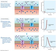
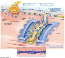
Muscle fibers maintain a resting membrane potential (typically around -90 mV) due to the distribution of sodium and potassium ions across the sarcolemma, maintained by the sodium-potassium pump.
Action Potentials
An action potential is a rapid, temporary change in membrane potential, involving depolarization (influx of Na+) and repolarization (efflux of K+). This electrical signal is propagated along the sarcolemma and T-tubules.
Neuromuscular Junction and Muscle Contraction
Neuromuscular Junction (NMJ)
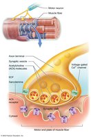
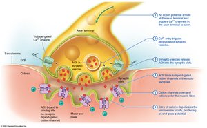
The NMJ is the synapse between a motor neuron and a skeletal muscle fiber, consisting of the axon terminal, synaptic cleft, and motor end plate. Acetylcholine (ACh) is the neurotransmitter that initiates muscle fiber depolarization.
Excitation-Contraction Coupling
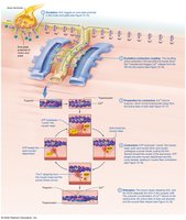
This process links the excitation of the sarcolemma to the contraction of the myofilaments via calcium release from the SR.
Contraction Phase: Crossbridge Cycle
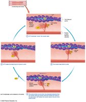
Calcium binds to troponin, causing tropomyosin to move and expose actin's active sites. Myosin heads bind to actin, undergo a power stroke, and detach upon ATP binding, repeating the cycle.
Muscle Relaxation
Relaxation occurs when ACh is degraded, calcium is pumped back into the SR, and tropomyosin blocks actin's active sites.
Rigor Mortis
After death, ATP is depleted, calcium remains in the cytosol, and myosin heads cannot detach from actin, causing muscles to stiffen until proteins degrade.

Energy Sources for Muscle Contraction
ATP Regeneration
Muscle fibers regenerate ATP through three main processes:
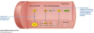
Creatine Phosphate Reaction: Provides immediate ATP for short bursts of activity.
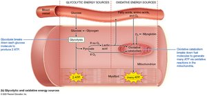
Glycolytic (Anaerobic) Catabolism: Produces ATP from glucose without oxygen; yields lactic acid.
Oxidative (Aerobic) Catabolism: Occurs in mitochondria, uses oxygen, and provides sustained ATP production.
Muscle Tension and Contraction Types
Muscle Twitch and Myogram
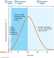
A muscle twitch is the response to a single action potential, recorded as a myogram with latent, contraction, and relaxation periods.
Wave Summation and Tetanus
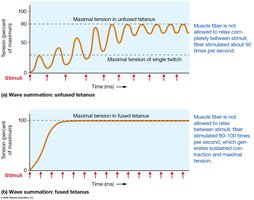
Repeated stimulation increases tension (wave summation). Unfused tetanus allows partial relaxation; fused tetanus produces sustained contraction.
Length-Tension Relationship
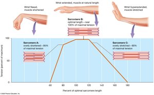
The amount of tension a muscle can produce depends on sarcomere length before contraction. Optimal overlap allows maximal crossbridge formation.
Types of Muscle Contractions
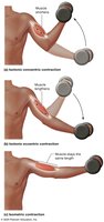
Isotonic Concentric: Muscle shortens as it contracts.
Isotonic Eccentric: Muscle lengthens while contracting.
Isometric: Muscle length does not change during contraction.
Muscle Fiber Types and Motor Units
Classes of Skeletal Muscle Fibers
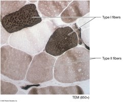
Slow-Twitch (Type I): Slow, fatigue-resistant, oxidative metabolism, high myoglobin (red muscle).
Fast-Twitch (Type II): Fast, powerful, fatigue quickly, glycolytic metabolism, low myoglobin (white muscle).
Motor Units
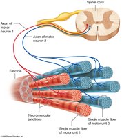
A motor unit consists of a motor neuron and all the muscle fibers it innervates. Recruitment of additional motor units increases force production.
Adaptations and Fatigue
Physical Training
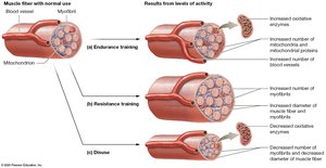
Endurance Training: Increases oxidative capacity and resistance to fatigue.
Resistance Training: Increases muscle fiber size (hypertrophy) and strength.
Disuse: Leads to atrophy and decreased strength/endurance.
Muscle Fatigue
Fatigue results from depletion of metabolites, decreased oxygen, accumulation of chemicals, and environmental factors. Recovery requires restoration of homeostasis and oxygen consumption (EPOC).
Smooth and Cardiac Muscle
Smooth Muscle
Smooth muscle is found in hollow organs and is responsible for peristalsis, sphincter formation, and regulation of flow. It lacks striations and contracts via a different mechanism involving calmodulin and myosin light-chain kinase.
Cardiac Muscle
Cardiac muscle is striated, branched, and connected by intercalated discs. It is autorhythmic due to pacemaker cells and contracts as a unit to pump blood.
