 Back
BackMuscle Tissue and Physiology: Structure, Function, and Key Terminology
Study Guide - Smart Notes
 Tailored notes based on your materials, expanded with key definitions, examples, and context.
Muscle Tissue and Physiology
Overview of Muscle Tissue
Muscle tissue is specialized for contraction and is responsible for movement of the body and its parts. There are three main types of muscle tissue: skeletal, cardiac, and smooth muscle. Each type has unique structural and functional characteristics.
Skeletal muscle: Voluntary, striated muscle attached to bones for movement.
Cardiac muscle: Involuntary, striated muscle found only in the heart.
Smooth muscle: Involuntary, non-striated muscle found in walls of hollow organs.
Structure of Skeletal Muscle
Muscle Fiber Organization
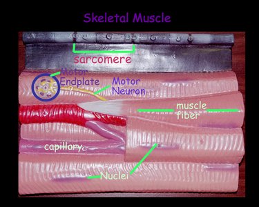
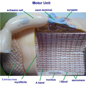
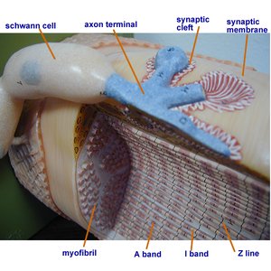
Skeletal muscle fibers are long, cylindrical cells containing multiple nuclei. Each fiber is composed of myofibrils, which are further divided into sarcomeres—the basic contractile units of muscle.
Myofibrils: Bundles of protein filaments (actin and myosin) responsible for contraction.
Sarcomere: The repeating unit between two Z lines; contains thick (myosin) and thin (actin, troponin, tropomyosin) filaments.
Connective tissue layers: Endomysium (surrounds each fiber), perimysium (surrounds fascicles), epimysium (surrounds entire muscle).
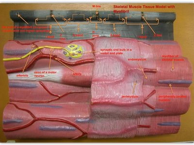
Sarcomere Structure
The sarcomere is the functional unit of muscle contraction. It is defined by the area between two Z disks and contains overlapping thick and thin filaments.
Z disk: Boundary of the sarcomere; anchors thin filaments.
M line: Center of the sarcomere; holds thick filaments together.
Thin filaments: Composed of actin, tropomyosin, and troponin.
Thick filaments: Composed of myosin molecules with projecting heads.

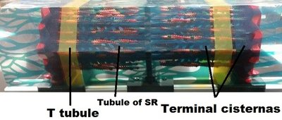
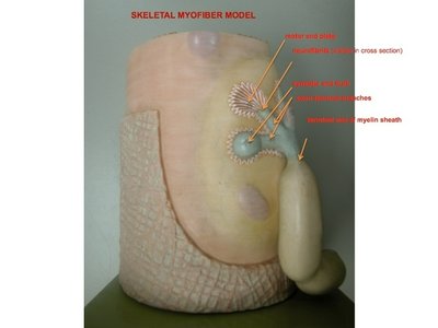
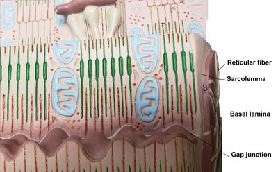
Transverse Tubules and Sarcoplasmic Reticulum
Muscle contraction is regulated by the release of calcium ions from the sarcoplasmic reticulum (SR), which is closely associated with transverse (T) tubules. T tubules conduct action potentials into the muscle fiber, triggering calcium release from the SR.
T tubules: Invaginations of the sarcolemma that transmit action potentials.
Sarcoplasmic reticulum: Specialized endoplasmic reticulum that stores calcium ions.
Terminal cisternae: Enlarged areas of the SR adjacent to T tubules; release calcium during contraction.
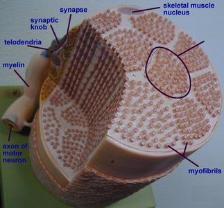
Neuromuscular Junction and Motor Unit
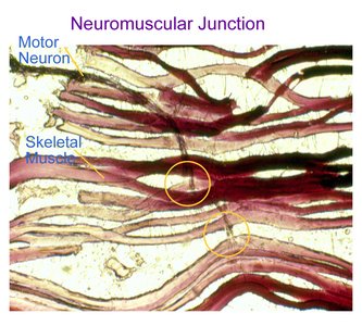
Neuromuscular Junction (NMJ)
The neuromuscular junction is the synapse between a motor neuron and a skeletal muscle fiber. It is the site where nerve impulses are transmitted to initiate muscle contraction.
Motor neuron: Nerve cell that transmits signals to muscle fibers.
Synaptic cleft: Small gap between the neuron and muscle fiber.
Motor endplate: Specialized region of the muscle fiber membrane at the NMJ.
Neurotransmitter: Acetylcholine (ACh) is released from the neuron to stimulate the muscle fiber.
Motor Unit
A motor unit consists of a single motor neuron and all the muscle fibers it innervates. The size of a motor unit determines the precision of muscle control.
Small motor units: Allow for fine control (e.g., eye muscles).
Large motor units: Allow for powerful contractions (e.g., thigh muscles).
Microscopic Anatomy of Muscle Fibers
Myofibrils and Associated Structures
Myofibrils are composed of repeating sarcomeres and are responsible for the striated appearance of skeletal muscle. Each myofibril contains thick and thin filaments arranged in a precise pattern.
Mitochondria: Provide ATP for muscle contraction.
Sarcoplasm: Cytoplasm of a muscle fiber, containing organelles and myofibrils.
Myofilaments: Actin (thin) and myosin (thick) filaments.
Muscle Terminology
Common Terms in Muscle Anatomy
Muscle names often reflect their location, shape, size, orientation, or function. Understanding these terms helps in identifying and describing muscles accurately.
Term | Meaning | Example |
|---|---|---|
Carpi | Wrist area | Extensor carpi ulnaris muscle |
Cervicis | Neck area | Splenius cervicis muscle |
Femoris | Thigh area | Quadriceps femoris muscle group |
Gluteus | Buttock area | Gluteus maximus muscle |
Oblique | At an angle | External oblique muscle |
Biceps | Two heads | Biceps brachii muscle |
Triceps | Three heads | Triceps brachii muscle |
Deltoid | Triangular shape | Deltoid muscle |
Maximus | Largest | Gluteus maximus muscle |
Minimus | Smallest | Gluteus minimus muscle |
Longus | Long | Adductor longus muscle |
Brevis | Short | Adductor brevis muscle |
Magnus | Large | Adductor magnus muscle |
Trapezius | Shaped like a trapezoid | Trapezius muscle |
Summary
This guide covers the essential structure and function of muscle tissue, focusing on skeletal muscle anatomy, the neuromuscular junction, and key terminology. Understanding these concepts is fundamental for further study in anatomy and physiology, especially regarding muscle contraction and movement.
