 Back
Muscle Tissue and Physiology: Structure, Function, and Mechanisms
Study Guide - Smart Notes
 Tailored notes based on your materials, expanded with key definitions, examples, and context.
Muscle Tissue Types
Overview of Muscle Tissue
Muscle tissue is a fundamental component of the human body, responsible for movement, posture, and various physiological functions. There are three primary types of muscle tissue: skeletal, cardiac, and smooth. Each type has distinct structural and functional characteristics.
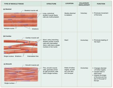
Skeletal Muscle: Long, cylindrical, multinucleated cells with striations; voluntary control; attached to bones; produces movement.
Cardiac Muscle: Short, branched cells with a single nucleus, striations, and intercalated discs; involuntary control; found in the heart; propels blood.
Smooth Muscle: Spindle-shaped cells with a single central nucleus; non-striated; involuntary control; lines hollow organs; propels substances and regulates diameter.
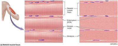
Skeletal Muscle Tissue
Skeletal muscle fibers are elongated, multinucleated cells with visible striations. They are mostly attached to the skeleton and are under voluntary control, enabling conscious movement.
Structure: Long, cylindrical fibers; multiple nuclei located peripherally; striated appearance.
Location: Attached to bones or skin.
Function: Voluntary movement, locomotion, manipulation, and expression.
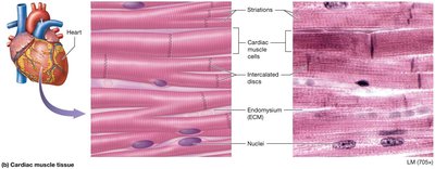
Cardiac Muscle Tissue
Cardiac muscle cells are shorter, branched, and typically have a single nucleus. They possess striations and intercalated discs, which facilitate synchronized contraction. Cardiac muscle is involuntary and found only in the heart.
Structure: Short, branched cells; single nucleus; striations; intercalated discs.
Location: Walls of the heart.
Function: Propels blood into circulation; involuntary control.
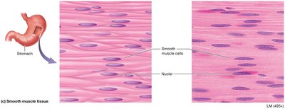
Smooth Muscle Tissue
Smooth muscle cells are spindle-shaped with a single, centrally located nucleus. They lack striations and are found in the walls of hollow organs, blood vessels, and other locations. Smooth muscle is involuntary and responsible for slow, sustained contractions.
Structure: Spindle-shaped cells; single central nucleus; non-striated.
Location: Walls of hollow organs (e.g., stomach, blood vessels).
Function: Propels substances; changes diameter of organs and vessels; involuntary control.
Structure of Muscle Cells
Muscle Cell (Myocyte) Anatomy
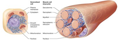
Muscle cells, or myocytes, contain specialized organelles and structures that facilitate contraction. The sarcoplasm is the cytoplasm of the muscle cell, and the sarcolemma is its plasma membrane. Myofibrils are bundles of proteins responsible for contraction, and the sarcoplasmic reticulum (SR) regulates calcium ion levels.
Sarcoplasm: Muscle cell cytoplasm.
Sarcolemma: Muscle cell plasma membrane.
Myofibrils: Bundles of contractile proteins.
Sarcoplasmic Reticulum: Modified smooth ER; stores and releases calcium ions.
Size and Shape of Skeletal Muscle Fibers
Skeletal muscle fibers are among the longest cells in the body, with a diameter of about 100 µm and lengths up to 30 cm. They are multinucleated and striated, reflecting their origin from fused myoblasts.

Organization of Skeletal Muscle
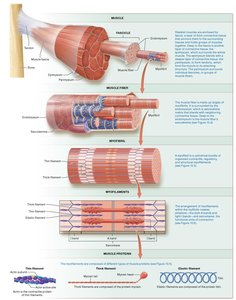
Skeletal muscle is organized into fibers, fascicles, and whole muscles, each surrounded by connective tissue layers:
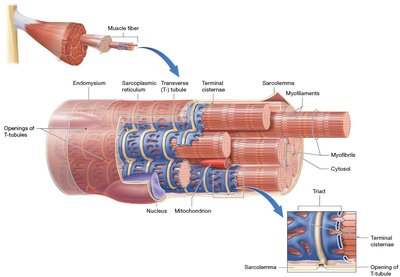
Endomysium: Surrounds each muscle fiber.
Perimysium: Surrounds each fascicle.
Epimysium: Covers the entire muscle.
Deep fascia: Binds muscles into functional groups.
Tendon: Attaches muscle to bone.
Aponeuroses: Flat, broad fibrous sheets.
Structure of Skeletal Muscle Fiber
The sarcolemma forms inward extensions called transverse tubules (T-tubules), which surround each myofibril and facilitate the conduction of nerve impulses. Terminal cisternae are enlarged portions of the SR flanking each T-tubule, forming a triad.
Myofibril and Myofilament Structure
Types of Myofilaments
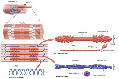
Myofibrils are composed of three types of myofilaments:
Thick Filaments: Composed of myosin; have heads that form cross-bridges during contraction.
Thin Filaments: Composed of actin, tropomyosin, troponin, and nebulin; actin has active sites for myosin binding.
Elastic Filaments: Composed of titin; provide elasticity and stability.
Sarcomere Structure and Banding
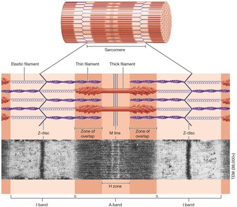
The sarcomere is the functional unit of muscle contraction, defined by the arrangement of myofilaments. It contains distinct bands and lines:
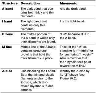
I band: Light region; only thin filaments.
A band: Dark region; thick and thin filaments.
H zone: Middle of A band; only thick filaments.
M line: Structural proteins holding thick filaments.
Z-disc: Anchors thin and elastic filaments; boundary of sarcomere.
Sarcomere Band Mnemonics
Structure | Description | Mnemonic |
|---|---|---|
A band | Dark band; thick and thin filaments | A is the dArk band |
I band | Light band; only thin filaments | I is the lIght band |
H zone | Middle of A band; only thick filaments | "Ha!" because H is in the A band |
M line | Middle line; structural proteins | "M" for middle/myosin |
Z-disc | Line bisecting I band; anchors filaments | "Z" shape |
Muscle Contraction Mechanism
Sliding-Filament Mechanism
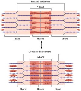
Muscle contraction occurs via the sliding-filament mechanism, where thin filaments slide past thick filaments, shortening the sarcomere and generating tension. The I bands and H zone narrow, while the A band remains unchanged.
Electrophysiology of Muscle Cells
Membrane Potential and Ion Gradients
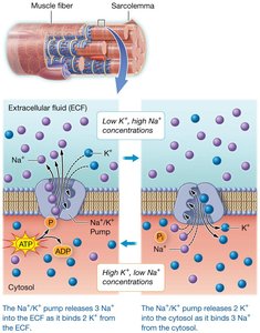
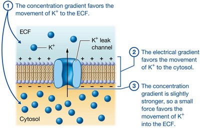
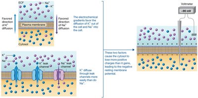
Muscle cells maintain a resting membrane potential through the action of ion gradients and channels. The sodium-potassium pump (Na+/K+ ATPase) moves three sodium ions out and two potassium ions in, creating a negative resting potential inside the cell.
Leak Channels: Always open; allow ions to follow gradients.
Gated Channels: Open in response to stimuli.
Electrochemical Gradient: Combination of concentration and electrical gradients determines ion movement.
Generation of Resting Membrane Potential
The resting membrane potential is established as potassium ions diffuse out of the cell more easily than sodium ions enter, resulting in a net loss of positive charge and a negative potential inside the cell.
Action Potentials
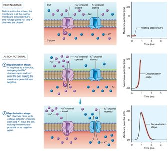
An action potential is a rapid, temporary change in membrane potential, initiated by the opening of voltage-gated sodium and potassium channels. It consists of two main stages:
Depolarization: Sodium channels open; sodium enters; membrane potential becomes less negative.
Repolarization: Sodium channels close; potassium channels open; potassium leaves; membrane potential returns to negative.
Neuromuscular Junction
Structure and Function
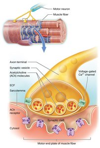
The neuromuscular junction (NMJ) is the synapse between a motor neuron and a muscle fiber. It consists of the axon terminal, synaptic cleft, and motor end plate. Acetylcholine (ACh) is the neurotransmitter released, which binds to receptors on the sarcolemma and initiates an action potential in the muscle fiber.
Axon Terminal: Contains synaptic vesicles with ACh.
Synaptic Cleft: Space between neuron and muscle; contains enzymes to degrade ACh.
Motor End Plate: Specialized region of sarcolemma with ACh receptors.
Summary Table: Muscle Tissue Types
Type | Structure | Location | Voluntary/Involuntary | Function |
|---|---|---|---|---|
Skeletal | Long, cylindrical, multinucleated, striated | Mostly attached to skeleton | Voluntary | Produces movement |
Cardiac | Short, branched, single nucleus, striated, intercalated discs | Heart | Involuntary | Propels blood |
Smooth | Spindle-shaped, single nucleus, non-striated | Walls of hollow organs | Involuntary | Propels substances, regulates diameter |
Additional info: Expanded explanations and context were added to ensure completeness and clarity for exam preparation.
