 Back
Muscle Tissue and Physiology: Structure, Function, and Contraction Mechanisms
Study Guide - Smart Notes
 Tailored notes based on your materials, expanded with key definitions, examples, and context.
Muscle Tissue Types
Overview of Muscle Tissue
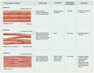
Muscle tissue is specialized for contraction and is essential for movement, posture, and various involuntary processes in the body. There are three main types of muscle tissue: skeletal, cardiac, and smooth muscle, each with distinct structures, locations, and functions.
Type of Muscle Tissue | Structure | Location | Voluntary/Involuntary | Function |
|---|---|---|---|---|
Skeletal | Long, cylindrical, striated muscle fibers; multinucleated | Mostly attached to skeleton | Voluntary | Produces movement of the body |
Cardiac | Short, wide, branching striated cells with intercalated discs; usually single nucleus | Heart | Involuntary | Produces beating of the heart |
Smooth | Thin, smooth muscle cells, single centrally located nucleus, no striations | Walls of hollow organs, eyes, skin, ducts of glands | Involuntary | Changes diameter of hollow organs, causes hair to stand erect, adjusts pupil size |
Phases of Skeletal Muscle Contraction
Excitation Phase
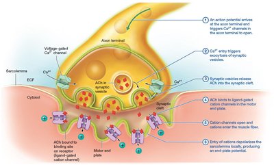
The excitation phase begins when a motor neuron stimulates a muscle fiber at the neuromuscular junction, leading to the generation of an action potential in the muscle cell membrane (sarcolemma).
Step 1: Action potential arrives at the axon terminal, opening voltage-gated Ca2+ channels.
Step 2: Ca2+ entry triggers exocytosis of synaptic vesicles containing acetylcholine (ACh).
Step 3: ACh diffuses across the synaptic cleft and binds to ligand-gated cation channels on the motor end plate.
Step 4: Cation channels open, allowing Na+ influx, depolarizing the sarcolemma and generating an end-plate potential.
Excitation-Contraction Coupling Phase
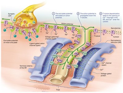
During excitation-contraction coupling, the action potential spreads along the sarcolemma and into the T-tubules, triggering the release of Ca2+ from the sarcoplasmic reticulum (SR).
Step 1: End-plate potential stimulates an action potential in the sarcolemma.
Step 2: Action potential propagates down T-tubules.
Step 3: T-tubule depolarization opens Ca2+ channels in the SR, releasing Ca2+ into the cytosol.
Preparation for Contraction
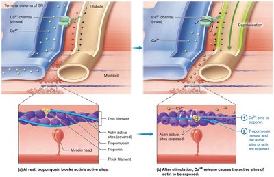
Ca2+ binds to troponin, causing tropomyosin to move and expose the active sites on actin, allowing myosin heads to bind and initiate contraction.
Step 1: Ca2+ binds to troponin.
Step 2: Tropomyosin shifts, exposing actin's active sites.
Contraction Phase: The Crossbridge Cycle
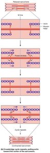
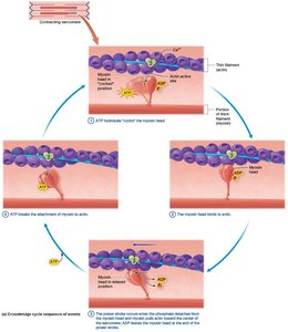
The crossbridge cycle is the mechanism by which myosin heads pull actin filaments, resulting in muscle contraction. This cycle repeats multiple times during a single contraction.
Step 1: ATP hydrolysis "cocks" the myosin head.
Step 2: Myosin head binds to actin, forming a crossbridge.
Step 3: Power stroke occurs as myosin pulls actin toward the M line; ADP is released.
Step 4: ATP binds to myosin, breaking the crossbridge and allowing the cycle to repeat.
Sliding-Filament Mechanism Analogy
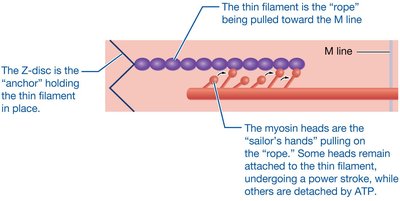
The sliding-filament mechanism can be compared to sailors pulling a rope (thin filament) toward an anchor (Z-disc). Some myosin heads are always attached, preventing the filament from sliding backward.
Muscle Tension at the Fiber Level
Muscle Twitch and Myogram
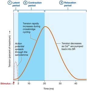
A muscle twitch is the response of a muscle fiber to a single action potential. It consists of three phases:
Latent Period: Time for action potential to spread and initiate contraction.
Contraction Period: Crossbridge cycling increases tension.
Relaxation Period: Ca2+ is pumped back into the SR, and tension decreases.
Motor Units and Recruitment
Motor Units
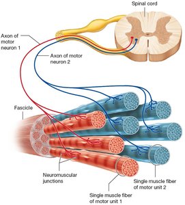
A motor unit consists of a single motor neuron and all the muscle fibers it innervates. The size of motor units varies depending on the precision required by the muscle.
Small motor units: Fine control (e.g., fingers)
Large motor units: Powerful contractions (e.g., back muscles)
Recruitment is the process of activating more motor units to increase muscle force.
Types of Muscle Contractions
Isotonic and Isometric Contractions
Muscle contractions can be classified based on changes in muscle length and tension:
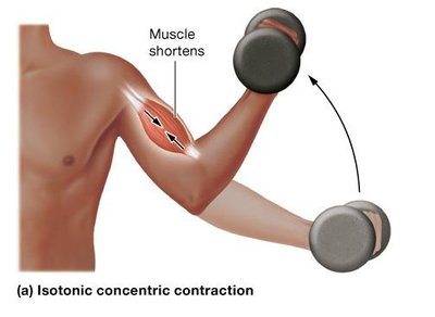
Isotonic Concentric Contraction: Muscle shortens as it generates force greater than the load (e.g., lifting a weight).
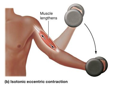
Isotonic Eccentric Contraction: Muscle lengthens as force generated is less than the load (e.g., lowering a weight).
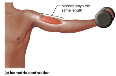
Isometric Contraction: Muscle length does not change (e.g., holding a weight stationary).
Muscle Relaxation
Mechanisms of Relaxation
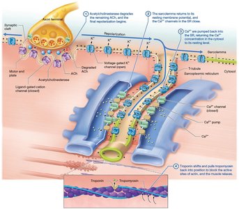
Muscle relaxation occurs when stimulation ceases, ACh is degraded, and Ca2+ is pumped back into the SR. Troponin and tropomyosin return to their resting positions, blocking actin's active sites.
Step 1: Acetylcholinesterase degrades ACh.
Step 2: Sarcolemma repolarizes; Ca2+ channels close.
Step 3: Ca2+ is pumped into the SR.
Step 4: Troponin and tropomyosin block actin's active sites; muscle relaxes.
Energy Sources for Skeletal Muscle
ATP and Muscle Contraction
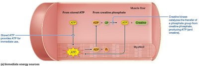
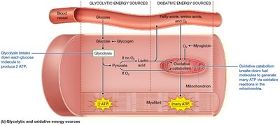
ATP is essential for muscle contraction and relaxation. Muscle fibers have three main mechanisms to regenerate ATP:
Immediate Source: Creatine phosphate donates a phosphate group to ADP to form ATP, catalyzed by creatine kinase. Equation:
Glycolytic (Anaerobic) Catabolism: Glucose is broken down in the cytosol to produce ATP and pyruvate. If oxygen is limited, pyruvate is converted to lactic acid. Equation:
Oxidative (Aerobic) Catabolism: Occurs in mitochondria, using oxygen to produce large amounts of ATP from glucose, fatty acids, or amino acids. Equation:
Additional info:
Myoglobin is an oxygen-binding protein in muscle fibers that stores and releases oxygen as needed for aerobic metabolism.
Creatine supplementation can enhance short-term, high-intensity muscle performance but has limited effects on endurance and may have health risks if taken in excess.
