Back
BackMuscle Tissue and Physiology: Structured Study Notes
Study Guide - Smart Notes
 Tailored notes based on your materials, expanded with key definitions, examples, and context.
Muscle Tissue and Physiology
Introduction to Muscle Tissue
Muscle tissue is a specialized tissue that contracts to produce movement and force. It is essential for various physiological functions, including locomotion, posture, and regulation of internal processes. Muscle cells possess unique properties that distinguish them from other cell types.
Muscular System Functions
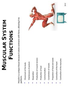
The muscular system performs several critical functions in the human body:
Movement of the body: Muscles contract to move bones and body parts.
Maintenance of posture: Muscles stabilize joints and maintain posture.
Respiration: Muscles such as the diaphragm facilitate breathing.
Production of heat: Muscle contractions generate heat, helping regulate body temperature.
Communication: Muscles enable facial expressions, speech, and gestures.
Constriction of vessels: Smooth muscle controls the diameter of blood vessels.
Regulation of food and heart: Muscles regulate movement of food through the digestive tract and contraction of the heart.
Contraction of the heart: Cardiac muscle contracts to pump blood.
Contraction of the eye: Muscles control pupil size and eye movement.
Types of Muscle Tissue
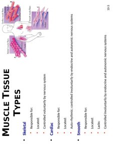
There are three main types of muscle tissue, each with distinct characteristics and functions:
Skeletal Muscle:
Responsible for voluntary movements.
Located attached to bones.
Controlled voluntarily by the nervous system.
Cardiac Muscle:
Responsible for pumping blood.
Located in the heart.
Auto-rhythmic, controlled involuntarily by endocrine and autonomic nervous systems.
Smooth Muscle:
Responsible for involuntary movements (e.g., constriction of vessels, movement of food).
Located in walls of hollow organs.
Controlled involuntarily by endocrine and autonomic nervous systems.
Properties of Muscle Cells
Muscle cells exhibit several unique properties that enable their function:
Contractility: Ability to contract and shorten, producing movement.
Excitability: Ability to respond to stimuli (chemical, mechanical, or electrical).
Conductivity: Ability to conduct electrical changes across the plasma membrane.
Extensibility: Ability to be stretched without being damaged.
Elasticity: Ability to return to original length after being stretched.
Skeletal Muscle
Structure and Function
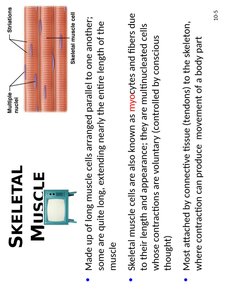
Skeletal muscle is composed of long, cylindrical cells called myocytes or muscle fibers. These cells are multinucleated and arranged parallel to one another. Skeletal muscle contractions are voluntary and are controlled by conscious thought.
Most skeletal muscles are attached to bones via connective tissue (tendons).
Contraction produces movement of body parts.
Skeletal Muscle Anatomy
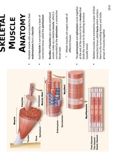
Skeletal muscle anatomy is organized into several layers of connective tissue:
Epimysium: Surrounds the entire muscle.
Perimysium: Surrounds bundles of muscle fibers called fascicles.
Endomysium: Surrounds individual muscle fibers.
Blood vessels and nerves penetrate these layers to supply the muscle tissue.
Myocytes: Specialized Terminology
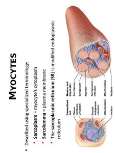
Muscle cells (myocytes) are described using specialized terminology:
Sarcoplasm: Cytoplasm of a muscle cell.
Sarcolemma: Plasma membrane of a muscle cell.
Sarcoplasmic reticulum (SR): Modified endoplasmic reticulum that stores and releases calcium ions.
Myocytes: Myofibrils and Proteins
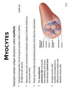
Myocytes contain unique structures called myofibrils, which are cylindrical organelles responsible for contraction:
Myofibrils are about one micrometer in diameter and make up 50-80% of cell volume.
They are composed of bundles of specialized proteins that allow for contraction.
The sarcoplasmic reticulum surrounds the myofibrils and stores calcium ions.
Skeletal Myocytes: Structure and Formation
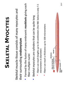
Skeletal muscle tissue consists of many myocytes and their surrounding endomysium. Myocytes are formed by the fusion of embryonic myoblasts, giving each fiber multiple nuclei. These cells can be quite long, reaching up to 30 centimeters in length and up to 100 micrometers in diameter.
Skeletal Myocyte Anatomy
Key anatomical features include:
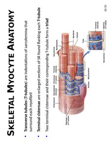
Transverse tubules (T-tubules): Invaginations of the sarcolemma that surround each myofibril.
Terminal cisternae: Enlarged sections of the sarcoplasmic reticulum flanking each T-tubule.
Two terminal cisternae and their corresponding T-tubule form a triad.
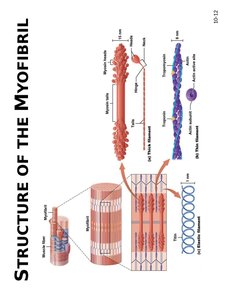
Structure of the Myofibril
Each myofibril is made of hundreds to thousands of proteins called myofilaments:
Contractile proteins: Generate tension (e.g., actin and myosin).
Regulatory proteins: Dictate when a fiber may contract (e.g., troponin, tropomyosin).
Structural proteins: Maintain proper alignment and stability (e.g., titin).
Myosin (Thick Filament)
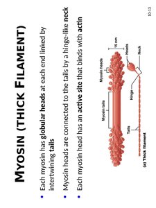
Myosin is a contractile protein forming the thick filament:
Each myosin has globular heads at each end linked by intertwining tails.
Heads are connected to tails by a hinge-like neck.
Each myosin head has an active site that binds with actin.
Actin (Thin Filament)
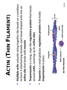
Actin is a contractile protein forming the thin filament:
Multiple actin subunits string together like beads on a necklace to form two intertwining strands.
Each bead-shaped actin has an active site that binds with myosin.
Troponin: A small globular regulatory protein that holds tropomyosin in place.
Tropomyosin: A long, rope-like regulatory protein that twists around actin, covering up its active sites.
Myofilaments Arranged in Sarcomeres
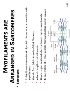
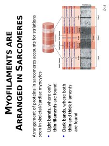
Myofilaments are organized into repeating units called sarcomeres:
Z disks: Serve as attachment for actin myofilaments.
I bands: From Z disks to ends of thick filaments.
A bands: Length of thick filaments.
H zone: Region in A band where actin and myosin do not overlap.
M line: Middle of H zone; delicate filaments holding myosin in place.
The Sliding-Filament Mechanism
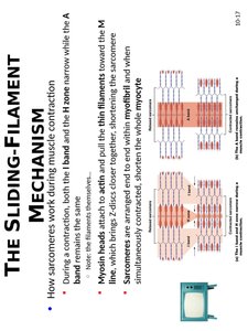
The sliding-filament mechanism explains how sarcomeres work during muscle contraction:
During contraction, both the I band and H zone narrow while the A band remains unchanged.
Myosin heads attach to actin and pull the thin filaments toward the M line, bringing Z disks closer together.
Sarcomeres are arranged end to end within myofibril and when simultaneously contracted, shorten the whole myocyte.
Muscle Contraction: Physiology
Membrane Potentials in Cells
Muscle contraction is initiated by changes in membrane potential:
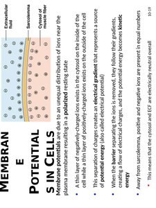
Membrane potential is due to an unequal distribution of ions near the plasma membrane, resulting in a polarized state.
This separation of charges creates an electrical gradient, representing a form of potential energy.
When the barrier separating the ions is removed, they flow down their gradients, creating a flow of electrical charges.
Measuring Membrane Potentials
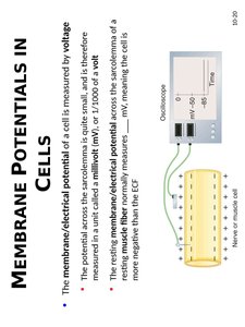
The membrane/electrical potential of a cell is measured by voltage. The potential across the sarcolemma is quite small and is measured in millivolts (mV).
The resting membrane/electrical potential across the sarcolemma of a resting muscle fiber is negative, meaning the cell is more negative than the extracellular fluid.
Na+ and K+ Concentration Gradients
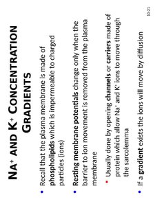
The plasma membrane is made of phospholipids, which are impermeable to charged particles (ions). Resting membrane potentials change only when the barrier to ion movement is removed from the plasma membrane, usually by opening channels or carriers.
Channels allow Na+ and K+ ions to move through the sarcolemma.
If a gradient exists, ions will move by diffusion.
Na+ and K+ Gradients in Muscle Cells
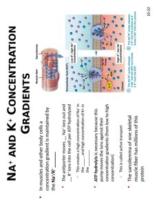
In muscle and other body cells, a concentration gradient is maintained by the Na+/K+ pump:
Na+ ions move out and K+ ions move in, creating a concentration gradient.
This is an example of active transport, which requires energy.
The sarcolemma of one skeletal muscle fiber has millions of these pumps.
Action Potentials
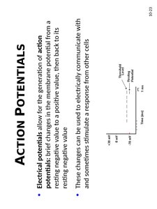
Electrical potentials allow for the generation of action potentials, brief changes in the membrane potential from a resting negative value to a positive value, then back to its resting value. These changes can be used to electrically communicate with and stimulate a response from other cells.
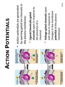
Action Potentials: Gated Ion Channels
Action potentials are generated by opening gated ion channels in the plasma membrane:
Ligand/chemically-gated channels: Open in response to the presence of a chemical.
Voltage-gated channels: Open in response to changes in the membrane potential.
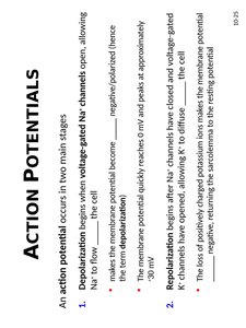
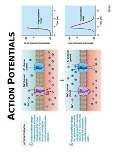
Action Potential Stages
An action potential occurs in two main stages:
Depolarization: Begins when voltage-gated Na+ channels open, allowing Na+ to flow into the cell, making the membrane potential become less negative (polarized).
Repolarization: Begins after voltage-gated Na+ channels have closed and voltage-gated K+ channels have opened, allowing K+ to diffuse out of the cell. The loss of positively charged potassium ions makes the membrane potential negative, returning the sarcolemma to the resting potential.
Action Potential Propagation
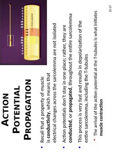
Action potentials are conducted, or propagated, throughout the entire sarcolemma. This process is very fast and results in depolarization of the entire sarcolemma, including the T-tubules. The arrival of the action potential at the T-tubules initiates muscle contraction.
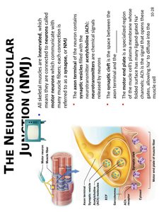
The Neuromuscular Junction (NMJ)
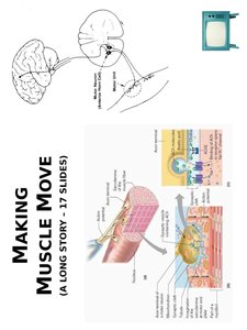
All skeletal myocytes are innervated, meaning they are connected to motor neurons at a synapse called the neuromuscular junction (NMJ):
Synaptic vesicles: Contain neurotransmitters (e.g., acetylcholine).
Synaptic cleft: Space between the neuron and muscle cell.
Motor end plate: Specialized region of the sarcolemma with ligand-gated Na+ channels.
Physiology of Skeletal Muscle Contraction
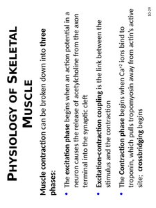
Muscle contraction can be broken down into three phases:
Excitation phase: Begins when an action potential in a neuron causes the release of acetylcholine from the axon terminal into the synaptic cleft.
Excitation-contraction coupling: The link between the stimulus and the contraction.
Contraction phase: Begins when Ca2+ ions bind to troponin, which pulls tropomyosin away from actin's active site, allowing crossbridging to begin.
Excitation Phase of Muscle Contraction
During the excitation phase, an action potential originating from the motor neuron arrives at the synaptic cleft, causing the release of acetylcholine. Acetylcholine binds to ligand-gated channels on the motor end plate, allowing Na+ ions to enter the muscle cell, generating an end-plate potential. This triggers an action potential in the sarcolemma, leading to muscle contraction.