 Back
Muscle Tissue and Physiology: Study Guide
Study Guide - Smart Notes
 Tailored notes based on your materials, expanded with key definitions, examples, and context.
Muscle Tissue and Physiology
Excitation-Contraction Coupling and ATP Generation
Muscle contraction is a complex process involving the interaction of electrical and chemical signals. The excitation-contraction coupling phase is especially dependent on the function of voltage-gated calcium channels in the sarcoplasmic reticulum. ATP generation in muscle cells varies depending on activity duration and intensity:
Excitation–contraction coupling: The process by which an action potential triggers calcium release, leading to muscle contraction.
ATP-generating mechanisms: After prolonged activity (e.g., marathon running), oxidative catabolism becomes the primary source of ATP.
Length-Tension Relationship
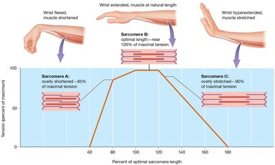
The tension a muscle fiber can produce depends on the initial length of its sarcomere. This relationship is crucial for understanding muscle function and optimal performance.
Optimal Length: The sarcomere length at which the maximum number of crossbridges can form, producing nearly 100% maximal tension.
Zone of Overlap: The degree of overlap between thick and thin filaments determines the ability of myosin heads to bind actin.
Overly Shortened Muscle: Excessive overlap limits tension production.
Overly Stretched Muscle: Insufficient overlap reduces tension.
Example: Wrist position affects sarcomere length and tension production.
Muscle Disorders
Several disorders affect muscle function:
Duchenne Muscular Dystrophy (DMD): Caused by a defective gene for dystrophin, leading to muscle degeneration and replacement by connective tissue.
Myasthenia Gravis: An autoimmune disorder where antibodies attack acetylcholine receptors, causing progressive skeletal muscle weakness.
Rigor Mortis: Postmortem muscle stiffening due to lack of ATP, preventing myosin head detachment from actin.
Tension Production: Timing and Frequency of Stimulation
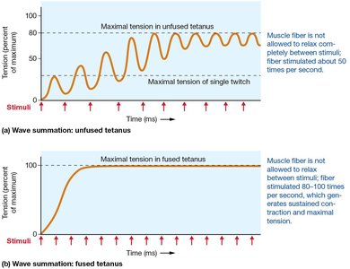
The amount of tension produced by a muscle fiber depends on the frequency of stimulation:
Wave Summation: Repeated stimulation increases tension as calcium accumulates.
Unfused (Incomplete) Tetanus: Partial relaxation between contractions; tension pulsates.
Fused (Complete) Tetanus: No relaxation; sustained maximal tension.
Muscle Toxins and Soreness
Tetanus: Caused by Clostridium tetani toxin, leading to tetanic spasms.
Botulism: Clostridium botulinum toxin blocks acetylcholine release, preventing contraction; used therapeutically as Botox.
Delayed-Onset Muscle Soreness (DOMS): Resulting from structural damage during eccentric contractions, not lactic acid.
Physical Training and Muscle Adaptation
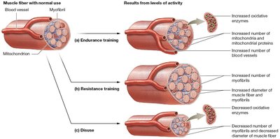
Muscle fibers adapt to different types of physical training:
Endurance Training: Increases oxidative enzymes, mitochondria, and blood vessels; improves resistance to fatigue.
Resistance Training: Increases myofibril number and fiber diameter (hypertrophy); decreases mitochondrial proteins and blood supply.
Disuse: Leads to atrophy, decreased oxidative enzymes, and reduced strength/endurance.
Muscle Fatigue and Recovery
Muscle fatigue is the inability to maintain exercise intensity, caused by:
Depletion of metabolites: Creatine phosphate, glycogen, and blood glucose.
Decreased oxygen availability: Reliance on glycolysis increases.
Chemical accumulation: Calcium, phosphate, and ADP interfere with contraction.
Environmental factors: Electrolyte disturbances from sweating.
Recovery Period: Excess Postexercise Oxygen Consumption (EPOC) corrects homeostatic disturbances, restores ion concentrations, and normalizes blood pH.
Smooth Muscle
Functions of Smooth Muscle
Smooth muscle is essential for various physiological functions:
Peristalsis: Rhythmic contractions propel materials through hollow organs.
Formation of sphincters: Rings of smooth muscle regulate passage of substances.
Regulation of flow: Alters diameter of blood vessels and airways.
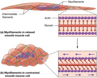
Structure of Smooth Muscle Cells
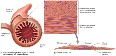
Smooth muscle cells differ structurally from skeletal and cardiac muscle:
No striations or sarcomeres: Actin filaments are obliquely arranged and anchored to dense bodies.
Intermediate filaments: Provide structural support.
Myosin heads: Found along the entire length of thick filaments.
Smooth Muscle Contraction and Relaxation
Smooth muscle contraction is triggered by various stimuli and involves unique mechanisms:
Calcium sources: Released from sarcoplasmic reticulum and enters from extracellular fluid.
Calmodulin: Calcium binds to calmodulin, activating myosin light-chain kinase (MLCK).
Crossbridge cycling: MLCK activates myosin ATPase, leading to contraction.
Latch state: Maintains tension with minimal ATP consumption.
Types of Smooth Muscle
Single-Unit (Unitary) Smooth Muscle: Cells linked by gap junctions contract as a unit; found in hollow organs and uterus.
Multi-Unit Smooth Muscle: Cells contract independently; found in eye muscles and arrector pili.
Cardiac Muscle
Structure and Function of Cardiac Muscle
Cardiac muscle cells are specialized for continuous, rhythmic contraction:
Striated with sarcomeres: Similar to skeletal muscle.
Intercalated discs: Connect cells via desmosomes and gap junctions, allowing coordinated contraction.
Autorhythmicity: Pacemaker cells generate action potentials spontaneously.
Functions of Cardiac Muscle
Automaticity: Contraction without neural stimulation.
Variable contraction tension: Controlled by the nervous system.
Extended contraction time: Longer than skeletal muscle.
Prevention of tetanus: Cardiac muscle cannot undergo wave summation or tetanic contractions.
Comparative Tables: Muscle Tissue Types
Structural and Functional Comparison
The following tables summarize key differences among skeletal, cardiac, and smooth muscle tissues:
Characteristic | Skeletal | Cardiac | Smooth |
|---|---|---|---|
Anatomical Location | Attached to bones or skin | Walls of the heart | GI, GU, respiratory, reproductive, blood vessels |
Histology | Single, very long, cylindrical, multinucleated, with obvious striations | Branching, chains of cells, uni- or binucleated, striations | Single, fusiform, uninucleated, lack striations |
Connective Tissue | Epimysium, perimysium, endomysium | Endomysium | Endomysium |
Presence of myofibrils | Yes | Yes, but irregular thickness | Presence of actin and myosin |
Sarcomere | Yes | Yes | No |
T tubules | Yes (triad) | Yes, larger diameter | No |
Sarcoplasmic reticulum | Yes, less than skeletal muscle with cistern terminal | Equivalent to cardiac | Equivalent to cardiac |
Gap junctions | No | Yes – intercalated discs | Yes – single-unit muscle |
Fibers exhibit individual neuromuscular junctions | Yes | No | No in single unit, yes in multiunit |
Regulation of contraction | Voluntary via somatic nervous system | Involuntary: intrinsic regulation, autonomic regulation, hormones, stretch | Involuntary: autonomic, hormones, local chemicals, stretch |
Characteristic | Skeletal | Cardiac | Smooth |
|---|---|---|---|
Sources for Ca2+ | Sarcoplasmic reticulum | SR and extracellular fluid | SR and extracellular fluid |
Site of calcium regulation | Troponin complex | Troponin complex | Calmodulin on myosin |
Presence of pacemaker | No | Yes | Yes – in single unit only |
Effect of nervous system stimulation | Excitation | Excitation or inhibition | Excitation or inhibition |
Speed of contraction | Slow to fast | Slow | Very slow |
Rhythmic contraction | No | Yes | Yes – single unit |
Response to stretch | Contractile strength increases with degree of stretch | Contractile strength increases with degree of stretch | Stress-relaxation response |
Respiration | Aerobic and anaerobic | Aerobic | Mainly anaerobic |
Additional info: These tables provide a concise comparison of muscle tissue types, aiding in understanding their unique properties and functions.
