 Back
BackMuscle Tissue and the Muscular System: Structured Study Notes
Study Guide - Smart Notes
 Tailored notes based on your materials, expanded with key definitions, examples, and context.
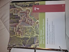
Muscle Tissue and the Muscular System
Overview of Muscle Tissue
The muscular system is essential for movement, posture, and vital bodily functions. Muscle tissue is specialized for contraction, enabling the body to move and maintain its structure. There are three main types of muscle tissue: skeletal, cardiac, and smooth.
Skeletal Muscle: Voluntary, striated muscle attached to bones; responsible for body movement.
Cardiac Muscle: Involuntary, striated muscle found only in the heart; responsible for pumping blood.
Smooth Muscle: Involuntary, non-striated muscle found in walls of internal organs; responsible for movements such as peristalsis.
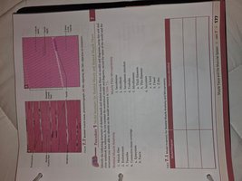
Microscopic Anatomy of Muscle Tissue
Muscle fibers are elongated cells containing multiple nuclei and specialized structures for contraction. The arrangement of actin and myosin filaments gives skeletal and cardiac muscle their striated appearance.
Myofibrils: Bundles of protein filaments within muscle fibers.
Sarcomere: The functional unit of muscle contraction, defined by the arrangement of actin and myosin.
Striations: Alternating light and dark bands seen in skeletal and cardiac muscle.
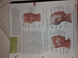
Major Muscles of the Head and Neck
The muscles of the head and neck are responsible for facial expressions, mastication, and movement of the head. Key muscles include the frontalis, temporalis, masseter, sternocleidomastoid, and others.
Frontalis: Raises eyebrows and wrinkles forehead.
Temporalis: Elevates and retracts the mandible.
Masseter: Main muscle for chewing.
Sternocleidomastoid: Rotates and flexes the neck.
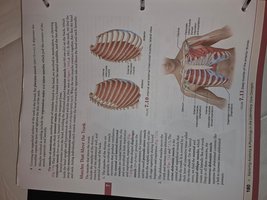
Muscles of the Thorax and Abdomen
These muscles are involved in respiration, posture, and movement of the trunk. The intercostal muscles assist with breathing, while the rectus abdominis, external oblique, and others stabilize the core.
Intercostal Muscles: Located between ribs; aid in breathing.
Rectus Abdominis: Flexes the vertebral column.
External Oblique: Rotates and laterally flexes the trunk.
Muscles of the Back
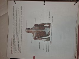
The muscles of the back support posture and allow for movement of the spine and shoulders. Major muscles include the trapezius, latissimus dorsi, and erector spinae.
Trapezius: Moves the scapula and supports the arm.
Latissimus Dorsi: Extends, adducts, and rotates the arm.
Erector Spinae: Extends and maintains posture of the vertebral column.
Muscles of the Upper Limb
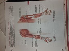
These muscles allow for movement of the shoulder, arm, forearm, and hand. Important muscles include the deltoid, biceps brachii, triceps brachii, flexor and extensor groups.
Deltoid: Abducts the arm.
Biceps Brachii: Flexes the elbow and supinates the forearm.
Triceps Brachii: Extends the elbow.
Muscles of the Lower Limb
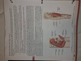
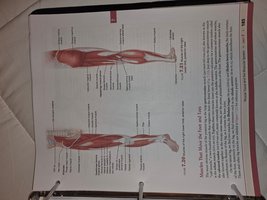
The muscles of the lower limb are responsible for movement and support of the hip, thigh, leg, and foot. Key muscles include the gluteus maximus, quadriceps femoris, hamstrings, gastrocnemius, and tibialis anterior.
Gluteus Maximus: Extends and laterally rotates the hip.
Quadriceps Femoris: Extends the knee.
Hamstrings: Flex the knee and extend the hip.
Gastrocnemius: Plantar flexes the foot.
Tibialis Anterior: Dorsiflexes the foot.
Muscle Attachments and Structure
Muscles attach to bones via tendons, which transmit the force of contraction to the skeleton. The origin is the fixed attachment, while the insertion moves with contraction.
Tendon: Connective tissue that attaches muscle to bone.
Origin: The stationary attachment point of the muscle.
Insertion: The movable attachment point.
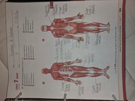
Summary Table: Major Muscles and Their Functions
The following table summarizes the major muscles discussed and their primary actions.
Muscle | Location | Primary Action |
|---|---|---|
Frontalis | Forehead | Raises eyebrows |
Masseter | Jaw | Chewing |
Sternocleidomastoid | Neck | Rotates/flexes neck |
Rectus Abdominis | Abdomen | Flexes vertebral column |
Deltoid | Shoulder | Abducts arm |
Biceps Brachii | Upper arm | Flexes elbow |
Quadriceps Femoris | Thigh | Extends knee |
Gastrocnemius | Calf | Plantar flexes foot |
Muscle Identification and Lab Exercises
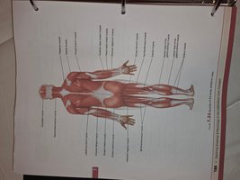
Students are often required to identify muscles on diagrams and models, and to understand their functions and locations. Practice exercises and quizzes reinforce this knowledge.
Labeling diagrams: Identifying muscles and their attachments.
Function matching: Associating muscles with their actions.
Lab exercises: Hands-on activities to reinforce anatomical knowledge.
Microscopic Structure of Skeletal Muscle
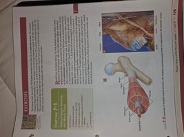
Skeletal muscle fibers are composed of repeating units called sarcomeres, which contain thick (myosin) and thin (actin) filaments. The sliding filament theory explains how muscles contract.
Sarcomere: The basic contractile unit of muscle fiber.
Actin and Myosin: Proteins responsible for muscle contraction.
Sliding Filament Theory: Muscle contraction occurs as actin and myosin filaments slide past each other.
Key Terms and Definitions
Muscle Fiber: A single muscle cell.
Fascicle: Bundle of muscle fibers.
Epimysium: Connective tissue surrounding the entire muscle.
Perimysium: Connective tissue surrounding fascicles.
Endomysium: Connective tissue surrounding individual muscle fibers.
Important Formulas and Concepts
Force Generation: The force a muscle generates depends on the number of fibers contracting and the frequency of stimulation.
Muscle Contraction Equation: (Force equals mass times acceleration)
Energy for Contraction: ATP is required for muscle contraction and relaxation.
Summary and Study Tips
Review diagrams and practice labeling major muscles.
Understand the microscopic structure and function of muscle tissue.
Memorize key muscle groups and their actions.
Apply knowledge in lab exercises and quizzes for reinforcement.
Additional info: Academic context was added to clarify muscle functions, microscopic anatomy, and lab exercises based on standard college-level anatomy and physiology curriculum.
