 Back
BackMuscle Tissue: Muscle Contraction and the Neuromuscular Junction
Study Guide - Smart Notes
 Tailored notes based on your materials, expanded with key definitions, examples, and context.
Muscle Tissue: Muscle Contraction
Overview of Skeletal Muscle Structure
Skeletal muscle fibers are specialized cells responsible for voluntary movement. Their structure is organized to facilitate contraction, which is the shortening of muscle fibers to produce force. - Sarcomere: The basic contractile unit of muscle, composed of actin (thin) and myosin (thick) filaments. - Sliding Filament Theory: Muscle contraction occurs when actin and myosin filaments slide past each other, shortening the sarcomere. 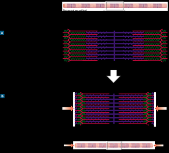
Sliding Filament Theory
The sliding filament theory explains how muscle fibers contract by the interaction of actin and myosin. - Actin: Thin filament, provides sites for myosin binding. - Myosin: Thick filament, uses ATP to bind and pull actin. - Contraction: Myosin heads attach to actin, pivot, and pull the filaments, shortening the sarcomere.
Neuromuscular Junction: Interaction of Neurons and Muscle Fibers
Neuron Structure
Neurons are specialized cells that transmit electrical signals. The axon terminal is the part of the neuron that interacts with muscle fibers. - Axon: Conducts electrical impulses to other cells. - Axon Terminal: Releases neurotransmitters to communicate with muscle fibers. 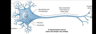
Structure and Function of the Neuromuscular Junction
The neuromuscular junction (NMJ) is where the axon terminal of a motor neuron meets the motor end plate of a muscle fiber. - Motor End Plate: Specialized region of the sarcolemma (muscle cell membrane) that receives signals. - Synaptic Cleft: Narrow space separating neuron and muscle fiber. 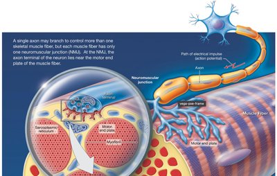
Events at the Neuromuscular Junction
The process of muscle contraction begins with a series of events at the NMJ:
ACh Storage: Acetylcholine (ACh), a neurotransmitter, is stored in vesicles in the axon terminal.
Action Potential Arrival: An electrical signal (action potential) arrives at the axon terminal.
ACh Release: The action potential triggers exocytosis, releasing ACh into the synaptic cleft.
ACh Binding: ACh binds to receptors on the motor end plate, opening channels for Na+ ions to enter the muscle fiber.
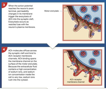
Muscle Fiber Action Potential: The influx of Na+ depolarizes the sarcolemma, generating an action potential in the muscle fiber. ACh is removed from the synaptic cleft by diffusion and breakdown by acetylcholinesterase (AChE).
Excitation-Contraction Coupling
Linking Action Potential to Muscle Contraction
Excitation-contraction coupling describes how the electrical signal in the muscle fiber leads to contraction. - Excitation: Action potential travels along the sarcolemma and into T tubules. - Calcium Release: Action potential triggers release of Ca2+ from the sarcoplasmic reticulum. 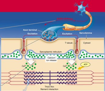
Muscle Contraction Cycle
The muscle contraction cycle consists of several steps involving actin and myosin:
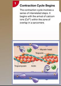
Ca2+ Binding: Calcium ions bind to troponin, causing tropomyosin to move and expose active sites on actin.
Cross-Bridge Formation: Myosin heads bind to exposed actin sites, forming cross-bridges.
Power Stroke: Myosin heads pivot, pulling actin filaments toward the M line and releasing ADP and phosphate.
Cross-Bridge Detachment: ATP binds to myosin, causing it to release actin.
Myosin Reactivation: Myosin hydrolyzes ATP, re-cocking its head for another cycle.
Key Structures in Muscle Contraction
- Sarcomere: Contains actin and myosin filaments. - Troponin and Tropomyosin: Regulatory proteins controlling access to actin's active sites. - ATP: Provides energy for myosin head movement and detachment.
Summary Table: Steps of Muscle Contraction Cycle
Step | Event | Key Molecules |
|---|---|---|
1 | Ca2+ released, binds to troponin | Ca2+, troponin, tropomyosin |
2 | Active sites exposed on actin | Actin, troponin, tropomyosin |
3 | Cross-bridge formation | Myosin, actin |
4 | Power stroke (myosin head pivots) | Myosin, ADP, Pi |
5 | Cross-bridge detachment | ATP, myosin, actin |
6 | Myosin reactivation | ATP, myosin |
Conditions for Continued Muscle Contraction
Muscle contraction continues as long as: - Ca2+ is available in the sarcoplasm. - ATP is available for myosin activity. - Sarcomere is not at maximum shortening limit (usually limited by joint movement).
Review: Muscle Contraction & Relaxation
Key Points for Exam Preparation
- Understand the events at the neuromuscular junction: axon terminal, motor end plate, ACh neurotransmitter, action potentials. - Know how action potentials initiate muscle contraction: T-tubules, Ca2+ release, contraction cycle. - Describe the muscle contraction cycle: actin and myosin interactions, sarcomere shortening. Example: During a biceps curl, motor neurons stimulate muscle fibers, leading to Ca2+ release and sarcomere shortening, resulting in arm flexion. Additional info: The process of muscle relaxation involves removal of Ca2+ from the sarcoplasm and breakdown of ACh, allowing tropomyosin to cover actin's active sites and ending contraction.
