 Back
BackMuscle Tissue Structure and Function: ANP Study Guide
Study Guide - Smart Notes
 Tailored notes based on your materials, expanded with key definitions, examples, and context.
Muscle Tissue Overview
Functions of Muscle Tissue
Muscle tissues are specialized for contraction, enabling movement, material transport, and heat generation. They respond to nervous system stimuli and are essential for bodily functions.
Movement: Muscles move parts of the body and materials through organs.
Heat Generation: Muscle contraction produces heat, helping maintain body temperature.
Posture and Stability: Muscles stabilize joints and maintain posture.
Properties of Muscle Tissue
Excitability, Contractility, Extensibility, Elasticity
Muscle cells possess four key properties that enable their function:
Excitability: Ability to respond to stimuli, usually electrical signals from nerves.
Contractility: Ability to shorten forcibly when stimulated.
Extensibility: Ability to be stretched beyond resting length.
Elasticity: Ability to return to original length after stretching or contracting.
Types of Muscle Tissue
Skeletal Muscle
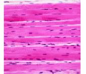
Skeletal muscle is attached to bones and is responsible for voluntary movements. It is striated and composed of long, multinucleated fibers.
Location: Attached to skeleton
Control: Voluntary
Appearance: Striated, cylindrical fibers
Function: Moves skeleton, maintains posture
Cardiac Muscle
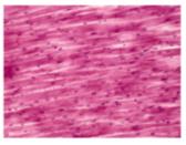
Cardiac muscle is found only in the heart wall. It is striated, branched, and under involuntary control. Cells are connected by intercalated discs, which allow coordinated contraction.
Location: Heart wall
Control: Involuntary
Appearance: Striated, branched fibers
Special Features: Intercalated discs, inherent rhythmicity
Smooth Muscle
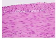
Smooth muscle is found in the walls of internal organs such as the intestines and blood vessels. It is non-striated and under involuntary control.
Location: Walls of hollow organs (e.g., intestines, blood vessels)
Control: Involuntary
Appearance: Spindle-shaped, non-striated fibers
Function: Moves materials through organs, regulates diameter of vessels
Microscopic Anatomy of Skeletal Muscle
Muscle Fiber Structure
Skeletal muscle fibers are long, cylindrical cells containing multiple nuclei. Each fiber is surrounded by a plasma membrane called the sarcolemma and contains myofibrils, which are bundles of actin and myosin filaments.
Sarcolemma: Plasma membrane of muscle cell
Sarcoplasm: Cytoplasm of muscle cell
Myofibrils: Rod-like bundles of actin and myosin
T tubules: Extensions of sarcolemma that carry electrical signals
Sarcoplasmic Reticulum: Stores and releases calcium ions
Sarcomere Structure
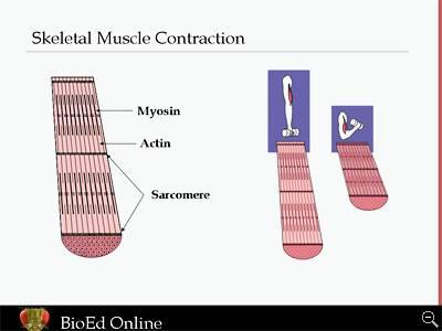
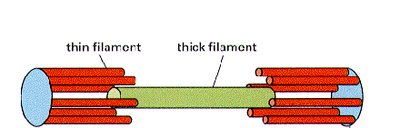
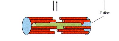
The sarcomere is the functional unit of muscle contraction, composed of repeating segments of actin (thin) and myosin (thick) filaments. The arrangement of these filaments gives skeletal muscle its striated appearance.
Z disc: Boundary of each sarcomere
A band: Region with thick (myosin) filaments
I band: Region with thin (actin) filaments
Titin: Elastic protein connecting Z disc to myosin, contributes to elasticity
Muscle Contraction Mechanism
Sliding Filament Theory
Muscle contraction occurs when actin and myosin filaments slide past each other, shortening the sarcomere and thus the muscle fiber. This process is powered by ATP and regulated by calcium ions.
Actin: Thin filament
Myosin: Thick filament
ATP: Provides energy for contraction
Calcium: Released from sarcoplasmic reticulum, triggers contraction
Muscle Attachments and Actions
Origin, Insertion, and Action
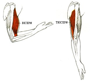
Muscles attach to bones at two points: the origin (fixed) and insertion (moved during contraction). The action is the movement produced by muscle contraction.
Origin: Attachment site that remains stationary
Insertion: Attachment site that moves
Action: Movement produced by contraction
Tendon: Dense regular connective tissue connecting muscle to bone
Muscle Organization and Control
Motor Units
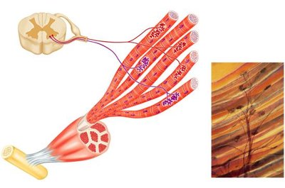
A motor unit consists of a motor neuron and all the muscle fibers it innervates. Activation of more motor units increases the force of contraction.
Fine Motor Control: Small muscles have fewer fibers per motor unit
Force Generation: Large muscles have more fibers per motor unit
Summary Table: Muscle Tissue Types
Type | Location | Control | Appearance | Function |
|---|---|---|---|---|
Skeletal | Bones | Voluntary | Striated, multinucleated | Movement, posture |
Cardiac | Heart wall | Involuntary | Striated, branched | Pumps blood |
Smooth | Internal organs | Involuntary | Non-striated, spindle-shaped | Moves materials |
Key Equations
Force of Muscle Contraction:
Sliding Filament Theory:
Additional info: The notes above expand on the original content by providing definitions, examples, and a summary table for clarity. Images included are directly relevant to the described tissue types, muscle structure, contraction mechanism, and motor unit organization.
