 Back
BackMuscle Tissue: Structure and Organization
Study Guide - Smart Notes
 Tailored notes based on your materials, expanded with key definitions, examples, and context.
Muscle Tissue
An Introduction to Muscle Tissue
Muscle tissue is a primary tissue in the human body, specialized for contraction and movement. There are three main types: skeletal muscle, cardiac muscle, and smooth muscle. Each type has unique structural and functional characteristics, but all share the ability to contract and generate force.
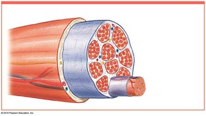
Organization of Skeletal Muscle
Structural Hierarchy of Skeletal Muscle
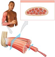
Skeletal muscle is organized in a hierarchical structure, from the whole muscle down to the microscopic myofibrils. This organization allows for efficient force generation and transmission.
Epimysium: The outermost layer of connective tissue surrounding the entire muscle.
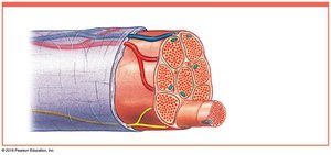
Perimysium: Surrounds bundles of muscle fibers called fascicles.
Endomysium: Surrounds individual muscle fibers (cells).
Tendon: Connective tissue that attaches muscle to bone.
Blood vessels and nerves are integrated throughout these layers to supply nutrients and control muscle activity.
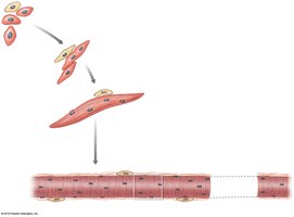
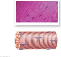
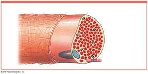
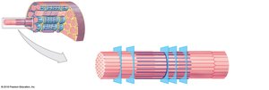
Development and Structure of Skeletal Muscle Fibers
Skeletal muscle fibers are large, multinucleate cells formed by the fusion of embryonic cells called myoblasts. These fibers contain hundreds of nuclei and are also known as striated muscle cells due to their banded appearance.
Myosatellite cells: Stem cells involved in muscle repair.
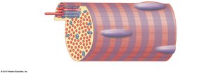
Sarcolemma: The plasma membrane of a muscle fiber.
Sarcoplasm: The cytoplasm of a muscle fiber.
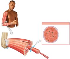
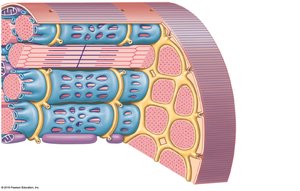
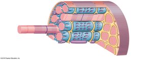
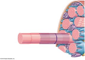
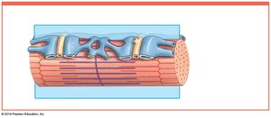
Internal Organization of Muscle Fibers
Within each muscle fiber, there are specialized structures that facilitate contraction:
Transverse tubules (T tubules): Invaginations of the sarcolemma that transmit action potentials into the cell.
Sarcoplasmic reticulum (SR): A network for storing and releasing calcium ions, essential for muscle contraction.
Myofibrils: Cylindrical structures composed of protein filaments (myofilaments) responsible for contraction.

Levels of Functional Organization
The functional organization of skeletal muscle can be summarized as follows:
Skeletal Muscle (organ): Surrounded by epimysium, contains muscle fascicles.
Muscle Fascicle: Surrounded by perimysium, contains muscle fibers.
Muscle Fiber: Surrounded by endomysium, contains myofibrils.
Myofibril: Surrounded by sarcoplasmic reticulum, consists of sarcomeres.
Sarcomere: The basic contractile unit, contains thick and thin filaments.
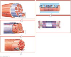
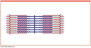
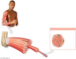
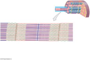
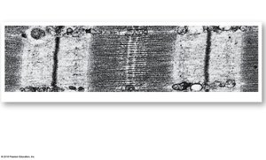
Sarcomere Structure
The sarcomere is the smallest functional unit of a muscle fiber. Its arrangement of thick (myosin) and thin (actin) filaments creates the striated appearance of skeletal muscle. Key components include:
A band: Dark band containing both thick and thin filaments.
I band: Light band containing only thin filaments.
H band: Region with only thick filaments.
Z line: Boundary between adjacent sarcomeres.
M line: Center of the A band, stabilizes thick filaments.
Titin: Elastic protein maintaining alignment and aiding in sarcomere recovery.

Summary Table: Levels of Skeletal Muscle Organization
Level | Surrounding Tissue | Contains |
|---|---|---|
Skeletal Muscle | Epimysium | Muscle Fascicles |
Muscle Fascicle | Perimysium | Muscle Fibers |
Muscle Fiber | Endomysium | Myofibrils |
Myofibril | Sarcoplasmic Reticulum | Sarcomeres |
Sarcomere | None | Thick & Thin Filaments |
Key Terms and Concepts
Excitability: Ability to respond to stimuli.
Contractility: Ability to shorten forcibly.
Extensibility: Ability to stretch.
Elasticity: Ability to recoil after stretching.
Example: The biceps brachii muscle contracts to flex the forearm, demonstrating excitability, contractility, extensibility, and elasticity.
Additional info: The images included above directly illustrate the hierarchical structure of skeletal muscle, the development of muscle fibers, and the internal organization necessary for contraction. These visual aids reinforce the anatomical concepts described in the text.
