 Back
BackMuscle Tissue: Structure, Function, and Contraction Mechanisms
Study Guide - Smart Notes
 Tailored notes based on your materials, expanded with key definitions, examples, and context.
Muscle Tissue Overview
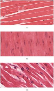
Types of Muscle Tissue
Muscle tissue is specialized for contraction and is essential for movement and support in the human body. There are three main types of muscle tissue:
Skeletal muscle: Voluntary, striated muscle attached to bones for movement.
Cardiac muscle: Involuntary, striated muscle found only in the heart.
Smooth muscle: Involuntary, non-striated muscle found in walls of hollow organs.
Functions of Skeletal Muscle
Skeletal muscles are responsible for a variety of essential functions:
Movement: Contraction of skeletal muscles moves the skeleton.
Posture: Muscles stabilize body position.
Thermogenesis: Muscle activity helps maintain body temperature.
Voluntary control: Enables conscious control over swallowing, defecation, and urination.
Support: Supports soft tissues, such as the abdominal wall.
Skeletal Muscle Structure
Connective Tissue Layers
Each skeletal muscle is organized into layers of connective tissue that provide structure and support:
Epimysium: Outermost layer; dense collagen fibers surround the entire muscle, separating it from other tissues.
Perimysium: Divides muscle into compartments called fascicles, each containing bundles of muscle fibers.
Endomysium: Surrounds individual muscle fibers within each fascicle; contains capillaries and nerve fibers.
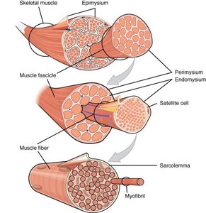
Muscle Fiber Anatomy
Skeletal muscle fibers are large, multinucleated cells with specialized structures:
Sarcolemma: The plasma membrane of a muscle fiber.
Sarcoplasm: The cytoplasm of a muscle fiber.
Sarcoplasmic reticulum (SR): Specialized smooth endoplasmic reticulum that stores calcium ions.
Myofibrils: Cylindrical structures composed of myofilaments (actin and myosin) responsible for contraction.
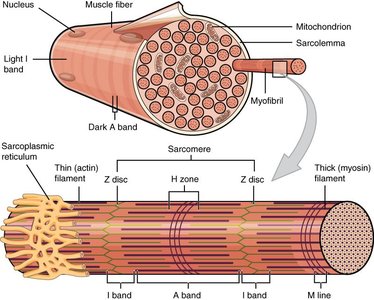
Organization of Myofibrils and Sarcomeres
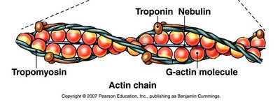
Myofibrils are made up of repeating units called sarcomeres, the functional units of muscle contraction. Sarcomeres contain:
Thick filaments: Composed of myosin.
Thin filaments: Composed of actin, troponin, and tropomyosin.
Regulatory proteins: Troponin and tropomyosin control access to actin's binding sites.

Neuromuscular Junction and Muscle Excitation
Neuromuscular Junction (NMJ)
The NMJ is the synapse where a motor neuron communicates with a skeletal muscle fiber to initiate contraction:
Motor neuron action potential arrives at the synaptic terminal.
Acetylcholine (ACh) is released into the synaptic cleft.
ACh binds to receptors on the motor end-plate of the sarcolemma, opening ion channels and generating a muscle action potential.
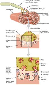
Excitation-Contraction Coupling
Excitation-contraction coupling links the action potential in the sarcolemma to muscle contraction:
Action potential travels along the sarcolemma and into the muscle fiber via T-tubules.
This triggers the release of Ca2+ from the sarcoplasmic reticulum (SR) into the sarcoplasm.
Calcium ions initiate contraction by binding to troponin.

Mechanism of Muscle Contraction
Role of Regulatory Proteins
Regulatory proteins control the interaction between actin and myosin:
Tropomyosin: Covers active sites on actin in resting muscle.
Troponin: Binds calcium ions, causing a conformational change that moves tropomyosin and exposes actin's active sites.
Cross-Bridge Cycle
The cross-bridge cycle describes the sequence of events during muscle contraction:
Calcium binds to troponin, exposing actin's active sites.
Myosin heads (already cocked with energy from ATP hydrolysis) bind to actin, forming cross-bridges.
Myosin heads pivot (power stroke), pulling actin filaments toward the center of the sarcomere and releasing ADP and Pi.
ATP binds to myosin, causing it to detach from actin.
ATP is hydrolyzed, re-cocking the myosin head for another cycle.

Sliding Filament Model
Muscle contraction is explained by the sliding filament model:
Thin filaments slide past thick filaments, shortening the sarcomere and the entire muscle fiber.
This process occurs simultaneously in all sarcomeres along a myofibril, resulting in muscle contraction.
Muscle Relaxation
Muscle relaxation occurs when:
Action potentials cease, and Ca2+ is actively pumped back into the SR.
Troponin and tropomyosin return to their resting positions, blocking actin's active sites.
Cross-bridge formation stops, and the muscle fiber returns to its resting length.
ATP and Muscle Metabolism
ATP Sources for Muscle Contraction
ATP is essential for both contraction and relaxation. Muscle fibers have three main pathways for ATP production:
Creatine phosphate pathway: Provides immediate ATP by transferring a phosphate group to ADP; lasts about 15 seconds.
Glycolysis: Anaerobic breakdown of glucose to produce ATP and pyruvate; yields 2 ATP per glucose molecule.
Aerobic respiration: Occurs in mitochondria when oxygen is available; each pyruvate yields about 17 ATP, making it the most efficient pathway.
Muscle Fatigue and Lactic Acid
During intense exercise, when oxygen is limited:
Glycolysis continues to provide ATP anaerobically.
Pyruvate is converted to lactic acid, which is transported to the liver for conversion back to glucose (Cori cycle).
Summary Table: Muscle Tissue Types
Muscle Type | Location | Control | Striations | Nuclei |
|---|---|---|---|---|
Skeletal | Attached to bones | Voluntary | Striated | Multinucleate |
Cardiac | Heart | Involuntary | Striated | Usually one |
Smooth | Walls of hollow organs | Involuntary | Non-striated | One |
Additional info: The above table summarizes the main distinguishing features of the three muscle tissue types for quick reference.
