 Back
BackMuscle Tissue: Structure, Function, and Contraction Mechanisms
Study Guide - Smart Notes
 Tailored notes based on your materials, expanded with key definitions, examples, and context.
Muscle Tissue
Introduction to Muscle Tissue
Muscle tissue is a primary tissue type in the human body, specialized for contraction and responsible for movement. There are three types of muscle tissue: skeletal muscle, cardiac muscle, and smooth muscle. Each type has distinct structural and functional characteristics.
Skeletal muscle: Moves the body by pulling on bones.
Cardiac muscle: Found only in the heart, controls the heartbeat.
Smooth muscle: Controls movements inside the body, such as in blood vessels and the digestive tract.
Functions and Properties of Muscle Tissue
Common Properties
All muscle tissues share several key properties:
Excitability: Ability to respond to stimuli.
Contractility: Ability to shorten and generate force.
Extensibility: Ability to be stretched without damage.
Elasticity: Ability to return to original length after stretching.
Functions of Skeletal Muscle
Producing movement
Maintaining posture and body position
Supporting soft tissues
Guarding body entrances and exits
Maintaining body temperature
Storing nutrients
Organization of Skeletal Muscle
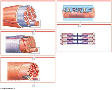
Structural Organization
Skeletal muscles are complex organs containing muscle tissue, connective tissues, blood vessels, and nerves. The connective tissue organization is hierarchical:
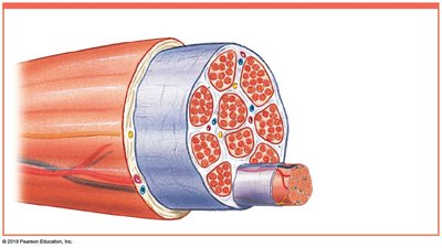
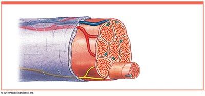
Epimysium: Surrounds the entire muscle; separates muscle from surrounding tissues.
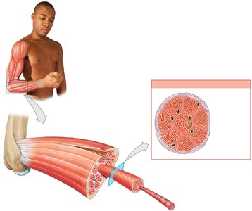
Perimysium: Surrounds bundles of muscle fibers called fascicles; contains blood vessels and nerves.
Endomysium: Surrounds individual muscle fibers; contains capillaries, myosatellite cells, and nerve fibers.
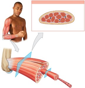
At the ends of muscles, collagen fibers from all three layers merge to form tendons (bundles) or aponeuroses (sheets), which attach muscles to bones.
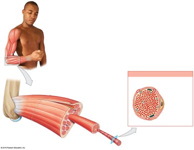
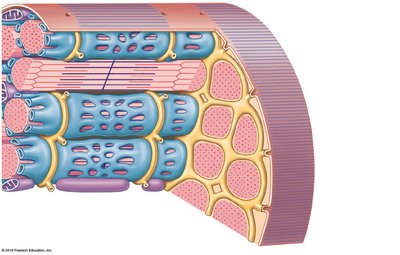
Skeletal Muscle Fibers
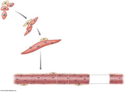
Development and Structure
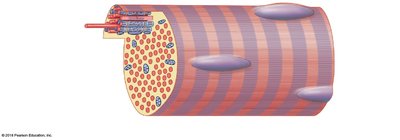
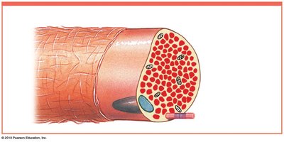
Skeletal muscle fibers are large, multinucleate cells formed by the fusion of embryonic cells called myoblasts. They are also known as striated muscle cells due to their banded appearance.
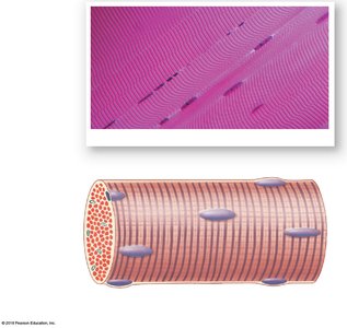
Sarcolemma: The plasma membrane of a muscle fiber.
Sarcoplasm: The cytoplasm of a muscle fiber.
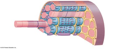
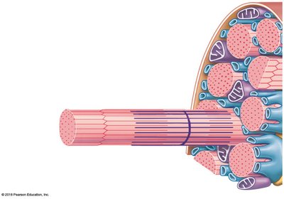
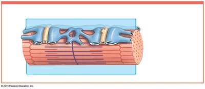
Transverse tubules (T tubules): Invaginations of the sarcolemma that transmit action potentials into the cell.
Sarcoplasmic reticulum (SR): Specialized endoplasmic reticulum that stores and releases calcium ions.
Myofibrils: Cylindrical structures within muscle fibers responsible for contraction, composed of myofilaments (actin and myosin).
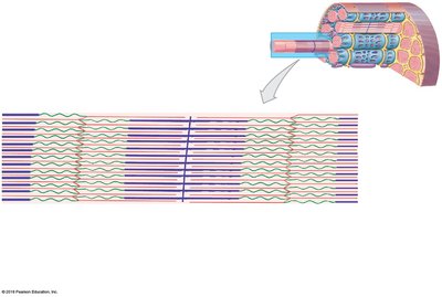
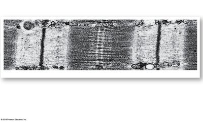
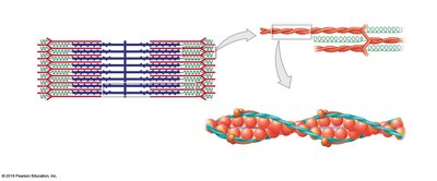
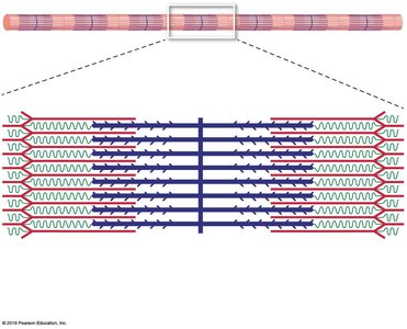
Sarcomere Structure
The sarcomere is the smallest functional unit of a muscle fiber. It is defined by the region between two Z lines and is responsible for the striated appearance of skeletal muscle.
A band: Dark region containing thick filaments (myosin).
I band: Light region containing only thin filaments (actin).
H band: Center of the A band with only thick filaments.
M line: Center of the H band; stabilizes thick filaments.
Z line: Boundary between adjacent sarcomeres; anchors thin filaments.
Titin: Elastic protein that helps restore sarcomere length after contraction.
Levels of Functional Organization
The organization of skeletal muscle can be summarized as follows:
Skeletal muscle (organ) → Muscle fascicles → Muscle fibers (cells) → Myofibrils → Sarcomeres → Myofilaments (actin and myosin)

Myofilament Structure
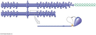
Thin filaments: Composed of F-actin (two rows of G-actin), nebulin, tropomyosin, and troponin.
Thick filaments: Composed of myosin molecules, each with a tail and two globular heads.
Mechanism of Muscle Contraction
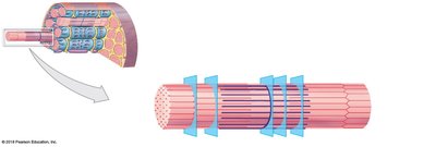
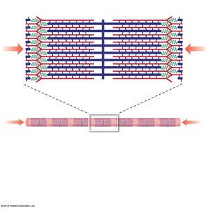
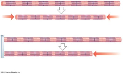
Sliding Filament Theory
Muscle contraction occurs when thin filaments slide past thick filaments, shortening the sarcomere without changing the length of the filaments themselves. Key observations:
H bands and I bands narrow.
Z lines move closer together.
A band width remains constant.
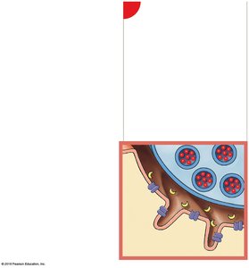
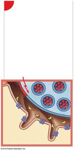
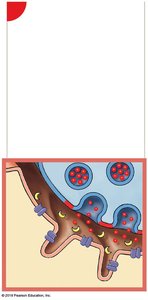
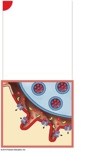
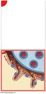
The Neuromuscular Junction (NMJ)
The NMJ is the synapse between a motor neuron and a skeletal muscle fiber. The process of muscle contraction initiation involves:
Action potential arrives at the axon terminal.
Acetylcholine (ACh) is released into the synaptic cleft.
ACh binds to receptors on the motor end plate, opening Na+ channels.
Na+ influx generates an action potential in the sarcolemma.
ACh is broken down by acetylcholinesterase (AChE).
Excitation-Contraction Coupling
After the action potential is generated in the sarcolemma, it travels down T tubules to the triads, triggering the release of Ca2+ from the SR. Ca2+ binds to troponin, causing the troponin-tropomyosin complex to shift and expose active sites on actin, initiating the contraction cycle.
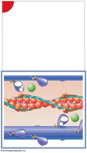
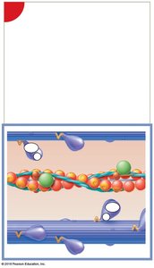
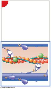
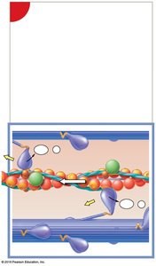
The Contraction Cycle
Contraction cycle begins (Ca2+ binds to troponin).
Active-site exposure (tropomyosin moves).
Cross-bridge formation (myosin binds to actin).
Myosin head pivoting (power stroke).
Cross-bridge detachment (ATP binds to myosin).
Myosin reactivation (ATP hydrolysis).
Muscle Relaxation
Relaxation occurs when neural stimulation ends, Ca2+ is pumped back into the SR, and active sites on actin are re-covered by tropomyosin. ATP is required for both contraction and relaxation.
Tension Production and Muscle Contractions
Tension Production
The amount of tension produced by a muscle fiber depends on:
Number of power strokes performed
Fiber's resting length at time of stimulation
Frequency of stimulation
Length-Tension Relationship
Maximum tension is produced when the maximum number of cross-bridges is formed, which occurs at an optimal sarcomere length.
Types of Muscle Contractions
Isotonic contractions: Muscle changes length (concentric: shortens; eccentric: lengthens).
Isometric contractions: Muscle develops tension but does not change length.
Energy for Muscle Contraction
ATP Production
Muscle fibers generate ATP through:
Direct phosphorylation of ADP by creatine phosphate (CP)
Anaerobic metabolism (glycolysis)
Aerobic metabolism (citric acid cycle and electron transport chain)
During rest, muscles store energy as glycogen and CP. During moderate activity, ATP is produced mainly by aerobic metabolism. At peak activity, glycolysis becomes the primary source, leading to lactate production.
Oxygen Debt and Recovery
After exercise, the body requires extra oxygen to restore normal metabolic conditions (EPOC). Lactate is recycled in the liver (Cori cycle).
Muscle Fiber Types and Performance
Types of Skeletal Muscle Fibers
Fast fibers: Large diameter, contract quickly, fatigue rapidly, few mitochondria.
Slow fibers: Small diameter, contract slowly, fatigue-resistant, many mitochondria, high myoglobin content.
Intermediate fibers: Characteristics between fast and slow fibers.
Muscle Hypertrophy and Atrophy
Hypertrophy: Increase in muscle size due to training.
Atrophy: Decrease in muscle size due to inactivity.
Cardiac and Smooth Muscle Tissue
Cardiac Muscle Tissue
Found only in the heart
Striated, branched cells with a single nucleus
Connected by intercalated discs (gap junctions and desmosomes)
Automaticity: Can contract without neural stimulation
Smooth Muscle Tissue
Found in walls of hollow organs
Non-striated, spindle-shaped cells with a single nucleus
Can contract over a wide range of lengths (plasticity)
Contraction regulated by neural, hormonal, or chemical factors
