 Back
BackMuscle Tissue: Structure, Function, and Contraction Mechanisms
Study Guide - Smart Notes
 Tailored notes based on your materials, expanded with key definitions, examples, and context.
Muscle Tissue
Introduction to Muscle Tissue
Muscle tissue is specialized for contraction and is essential for movement, posture, and various physiological processes. There are three main types of muscle tissue: skeletal, cardiac, and smooth, each with distinct structural and functional characteristics.
Skeletal Muscle: Voluntary, striated muscle attached to bones, responsible for body movement.
Cardiac Muscle: Involuntary, striated muscle found only in the heart, responsible for pumping blood.
Smooth Muscle: Involuntary, non-striated muscle found in the walls of hollow organs, responsible for moving substances through internal passageways.

Special Characteristics of Muscle Tissue
Excitability: Ability to receive and respond to stimuli.
Contractility: Ability to shorten when stimulated.
Extensibility: Ability to be stretched.
Elasticity: Ability to recoil to resting length after stretching.
Functions of Muscle Tissue
Movement: Skeletal muscles move the skeleton; cardiac muscle moves blood; smooth muscle moves substances through organs.
Posture and Support: Maintains body position and supports soft tissues.
Guarding Openings: Controls entrances and exits of digestive, urinary, and respiratory tracts.
Thermogenesis: Muscle contractions produce heat, helping maintain body temperature.
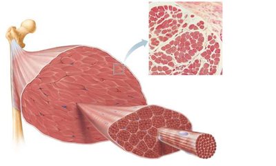
Skeletal Muscle Structure
Hierarchical Organization
Skeletal muscle is organized into several levels, each surrounded by connective tissue sheaths:
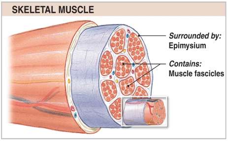
Muscle (organ): Surrounded by epimysium, contains bundles of fascicles.
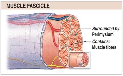
Fascicle: Surrounded by perimysium, contains bundles of muscle fibers.
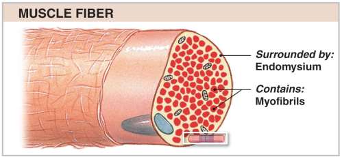
Muscle Fiber (cell): Surrounded by endomysium, contains myofibrils.
Myofibril: Composed of repeating units called sarcomeres.
Sarcomere: The functional contractile unit, composed of myofilaments (actin and myosin).
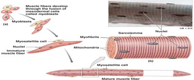
Development and Regeneration
Muscle fibers develop from the fusion of mesodermal cells called myoblasts. Myosatellite cells assist in muscle repair and regeneration.
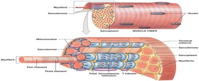
Internal Organization of a Muscle Fiber
Sarcolemma: The plasma membrane of a muscle fiber, conducts action potentials.
Sarcoplasm: The cytoplasm, containing myofibrils, mitochondria, glycogen, and myoglobin.
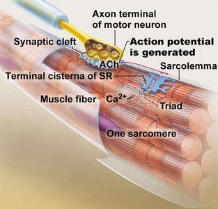
Sarcoplasmic Reticulum (SR): Specialized smooth ER that stores and releases calcium ions.
Transverse Tubules (T-tubules): Invaginations of the sarcolemma that transmit action potentials deep into the fiber.
Triad: A T-tubule flanked by two terminal cisternae of the SR, essential for excitation-contraction coupling.
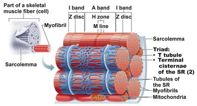
Sarcomere Structure and Function
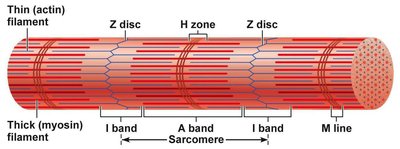
Sarcomere Organization
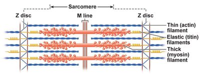
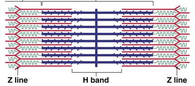
The sarcomere is the smallest contractile unit of muscle, defined as the region between two Z discs. It contains overlapping thick (myosin) and thin (actin) filaments, which create the striated appearance of skeletal muscle.
A Band: Dark region containing thick filaments; does not change length during contraction.
I Band: Light region containing only thin filaments; shortens during contraction.
H Zone: Central region with only thick filaments; shortens/disappears during contraction.
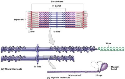
M Line: Center of the H zone; anchors thick filaments.
Z Disc: Boundary of the sarcomere; anchors thin filaments.
Myofilaments
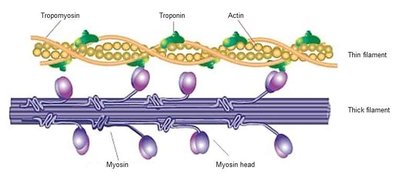
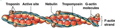
Thin Filaments: Composed mainly of actin, with regulatory proteins tropomyosin and troponin, and structural protein nebulin.
Thick Filaments: Composed of myosin molecules, each with a head (binds actin and ATP) and tail (toward M line). Structural proteins include titin and myomesin.
Muscle Contraction Mechanism
Neuromuscular Junction and Excitation
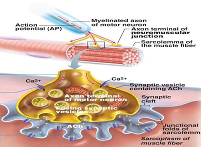
Muscle contraction is initiated by an action potential from a motor neuron, which triggers the release of acetylcholine (ACh) at the neuromuscular junction. ACh binds to receptors on the sarcolemma, causing depolarization and the generation of a muscle action potential.
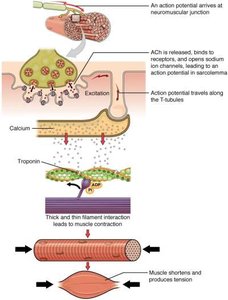
Excitation-Contraction Coupling
The action potential travels along the sarcolemma and down the T-tubules, triggering the release of Ca2+ from the sarcoplasmic reticulum. Calcium binds to troponin, causing a conformational change that moves tropomyosin and exposes myosin-binding sites on actin.
Cross-Bridge Cycle
The cross-bridge cycle describes the interaction between actin and myosin that leads to muscle contraction:
ATP Hydrolysis: Myosin head hydrolyzes ATP, becoming energized.
Cross-Bridge Formation: Energized myosin head binds to actin.
Power Stroke: Myosin head pivots, pulling actin toward the M line; ADP and Pi are released.
Cross-Bridge Detachment: New ATP binds to myosin, causing detachment from actin.
Reactivation: ATP is hydrolyzed, re-cocking the myosin head.
Sliding Filament Model
During contraction, thin filaments slide past thick filaments, shortening the sarcomere without changing the length of the filaments themselves. The I band and H zone shorten, while the A band remains constant.

Muscle Fiber Types and Adaptations
Types of Skeletal Muscle Fibers
Type | Contraction Speed | ATP Pathway | Fatigue Resistance | Myoglobin Content |
|---|---|---|---|---|
Slow Oxidative (Type I) | Slow | Aerobic | High | High |
Fast Oxidative (Type IIa) | Fast | Aerobic | Intermediate | Intermediate |
Fast Glycolytic (Type IIb) | Very Fast | Anaerobic | Low | Low |
Muscle Adaptations
Hypertrophy: Increase in muscle size due to resistance training (more myofibrils, mitochondria, and glycogen).
Atrophy: Decrease in muscle size due to inactivity.
Cardiac and Smooth Muscle Tissue
Cardiac Muscle
Found only in the heart; cells are striated, branched, and connected by intercalated discs (containing desmosomes and gap junctions).
Exhibits automaticity (can contract without neural stimulation).
Smooth Muscle
Found in walls of hollow organs; cells are spindle-shaped, non-striated, and have a single nucleus.
Responsible for involuntary movements such as peristalsis and regulation of vessel diameter.
Summary Table: Muscle Tissue Types
Characteristic | Skeletal | Cardiac | Smooth |
|---|---|---|---|
Body Location | Attached to bones or skin | Walls of the heart | Walls of hollow organs |
Cell Shape & Appearance | Long, cylindrical, multinucleate, striated | Branching, uni- or binucleate, striated | Spindle-shaped, uninucleate, no striations |
Key Equations
ATP Hydrolysis:
Creatine Phosphate Reaction:
Aerobic Respiration (simplified):
Anaerobic Glycolysis:
Additional info: This guide integrates foundational concepts of muscle tissue structure and function, excitation-contraction coupling, and the physiological basis of muscle contraction, as well as the differences between muscle tissue types. It is suitable for ANP college-level exam preparation.
