 Back
BackMuscle Tissue: Structure, Function, and Mechanisms
Study Guide - Smart Notes
 Tailored notes based on your materials, expanded with key definitions, examples, and context.
Muscle Tissue Overview
Introduction
Muscle tissue is a fundamental component of the human body, accounting for nearly half of its mass. It is specialized to transform chemical energy (ATP) into mechanical energy, enabling force generation and movement. The study of muscle tissue encompasses its types, characteristics, and functions.
Terminology: Prefixes such as myo, mys, and sarco are commonly used in muscle-related terms (e.g., sarcoplasm refers to muscle cell cytoplasm).
Types of Muscle Tissue:
Skeletal Muscle: Voluntary, striated, attached to bones and skin.
Cardiac Muscle: Involuntary, striated, found only in the heart.
Smooth Muscle: Involuntary, non-striated, found in walls of hollow organs.
Only skeletal and smooth muscle cells are elongated and referred to as muscle fibers.
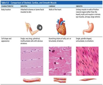
Comparison of Skeletal, Cardiac, and Smooth Muscle
The three types of muscle tissue differ in location, structure, and function.
Characteristic | Skeletal | Cardiac | Smooth |
|---|---|---|---|
Body Location | Attached to bones or skin | Walls of the heart | Walls of hollow organs (e.g., stomach, bladder) |
Cell Shape & Appearance | Long, cylindrical, multinucleate, striated | Branching chains, uni- or binucleate, striated | Spindle-shaped, uninucleate, non-striated |
Characteristics of Muscle Tissue
Four Main Properties
All muscle tissues share four essential characteristics:
Excitability (Responsiveness): Ability to receive and respond to stimuli.
Contractility: Ability to shorten forcibly when stimulated.
Extensibility: Ability to be stretched.
Elasticity: Ability to recoil to resting length after stretching.
Functions of Muscle Tissue
Key Functions
Muscle tissue performs several vital functions:
Produce Movement: Responsible for locomotion and manipulation (e.g., walking, digesting, pumping blood).
Maintain Posture and Body Position: Stabilizes the body during activity.
Stabilize Joints: Reinforces and supports joints.
Generate Heat: Muscle contraction produces heat, helping maintain body temperature.
Skeletal Muscle Anatomy
Structural Organization
Skeletal muscle is an organ composed of multiple tissue types and features:
Nerve and Blood Supply: Each muscle receives a nerve, artery, and veins.
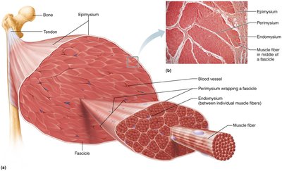
Connective Tissue Sheaths: Muscles and muscle fibers are covered by connective tissue for support and reinforcement.
Epimysium: Surrounds the entire muscle.
Perimysium: Surrounds fascicles (bundles of muscle fibers).
Endomysium: Surrounds individual muscle fibers.
Attachments:
Direct (fleshy): Epimysium fused to periosteum of bone or perichondrium of cartilage.
Indirect: Connective tissue wrappings extend beyond muscle as tendons or aponeuroses.
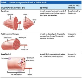
Structure & Organizational Levels of Skeletal Muscle
Skeletal muscle is organized hierarchically from the muscle organ to the muscle fiber.
Level | Description | Connective Tissue Wrapping |
|---|---|---|
Muscle (organ) | Hundreds to thousands of muscle cells, connective tissue, blood vessels, nerve fibers | Epimysium |
Fascicle (portion of muscle) | Discrete bundle of muscle cells, segregated from rest of muscle | Perimysium |
Muscle fiber (cell) | Elongated multinucleate cell, banded appearance | Endomysium |
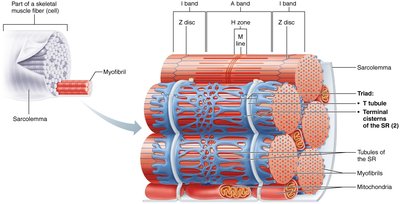
Muscle Fiber Microanatomy
Cellular Structure
Skeletal muscle fibers are long, cylindrical cells with multiple nuclei.
Sarcolemma: Plasma membrane of the muscle fiber.
Sarcoplasm: Cytoplasm of the muscle fiber, rich in glycosomes (glycogen storage) and myoglobin (oxygen storage).
Modified Organelles:
Myofibrils: Densely packed, rodlike elements responsible for muscle contraction.
Sarcoplasmic Reticulum: Specialized endoplasmic reticulum for calcium storage and release.
T Tubules: Extensions of the sarcolemma that conduct electrical impulses deep into the fiber.
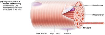
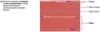
Myofibrils: Striations
Striations are alternating dark and light bands along the length of each myofibril, resulting from the arrangement of myofilaments.
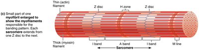
Myofibrils: Sarcomere
The sarcomere is the smallest contractile unit of muscle fiber, defined as the region between two Z discs. It contains an A band with half of an I band at each end.
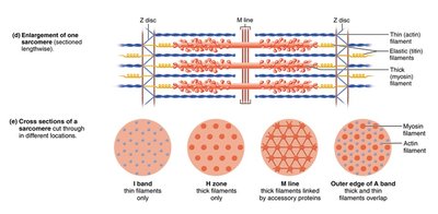
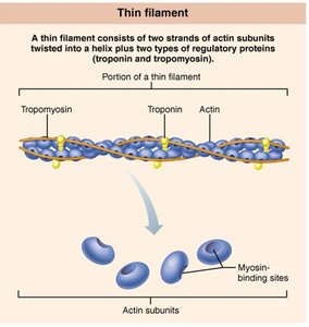
Myofibrils: Myofilaments
Myofilaments are the contractile proteins within the sarcomere, arranged in an orderly manner:
Actin (thin filaments): Composed of globular G actin subunits, each with a myosin-binding site.
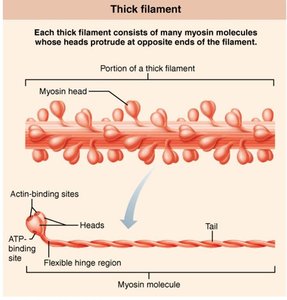
Myosin (thick filaments): Composed of myosin molecules with heads that bind to actin.
Tropomyosin and Troponin: Regulatory proteins bound to actin.
Titin (elastic filament): Maintains structure and elasticity of the sarcomere.
Sarcoplasmic Reticulum and T Tubules
Structure and Function
Sarcoplasmic Reticulum (SR): Network of smooth endoplasmic reticulum tubules surrounding each myofibril, regulates intracellular Ca2+ levels.
T Tubules: Invaginations of the sarcolemma that allow electrical impulses to reach deep into the muscle fiber.
Triad: Area formed by a T tubule and two terminal cisterns of the SR, crucial for excitation-contraction coupling.
Sliding Filament Model of Contraction
Mechanism of Muscle Contraction
The sliding filament model explains how muscles contract:
During contraction, thin filaments slide past thick filaments, increasing overlap.
Myosin heads bind to actin, forming cross bridges, and pull thin filaments toward the center of the sarcomere.
Neither filament changes length; the sarcomere shortens.


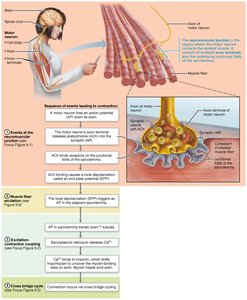
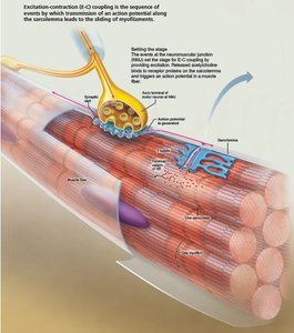
Muscle Fiber Contraction: The Big Picture
Overview of Contraction Steps
Muscle contraction is initiated by signals from the brain, transmitted via motor neurons. Four steps are required for skeletal muscle contraction:
Events at the neuromuscular junction
Muscle fiber excitation
Excitation-contraction coupling
Cross bridge cycling
Anatomy of Motor Neurons & Neuromuscular Junction (NMJ)
Structure and Function
Skeletal muscles are stimulated by somatic motor neurons.
Axons branch and form neuromuscular junctions with muscle fibers.
Axon terminal and muscle fiber are separated by the synaptic cleft.
Synaptic vesicles contain acetylcholine (ACh), which binds to receptors on the sarcolemma.
NMJ consists of axon terminals, synaptic cleft, and junctional folds.
Skeletal Muscle Contraction: Events at the Neuromuscular Junction
Stepwise Process
Action potential arrives at axon terminal.
Voltage-gated calcium channels open; calcium enters motor neuron.
Calcium triggers release of ACh into synaptic cleft.
ACh binds to receptors, opening Na+ channels and generating end plate potential.
Acetylcholinesterase degrades ACh, terminating the signal.
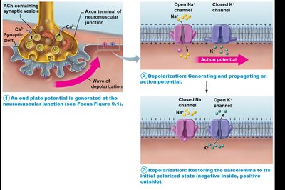
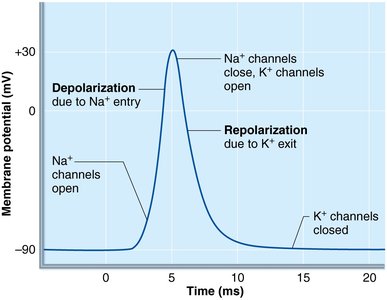
Muscle Fiber Excitation
Action Potential Generation
Resting sarcolemma is polarized (negative inside, positive outside).
Action potential is generated by changes in electrical charges:
Generation of end plate potential
Depolarization
Repolarization
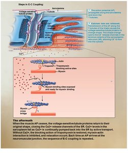
Excitation-Contraction (E-C) Coupling
Linking Electrical and Mechanical Events
AP propagates along sarcolemma and into T tubules.
Voltage-sensitive proteins trigger Ca2+ release from SR.
Ca2+ initiates contraction by enabling cross bridge formation.
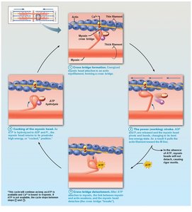
Cross Bridge Cycling
Mechanism of Force Generation
Cross bridge formation: Myosin head binds to actin.
Power stroke: Myosin head pivots, pulling actin toward M line.
Cross bridge detachment: ATP binds to myosin, causing detachment.
Cocking of myosin head: ATP hydrolysis re-energizes myosin head.
Clinical – Homeostatic Imbalance: Rigor Mortis
Postmortem Muscle Stiffening
Occurs 3–4 hours after death, peaks at 12 hours.
ATP production ceases, Ca2+ accumulates, cross bridges form and cannot detach.
Muscles remain contracted until proteins break down.
References
Marieb, E. N., & Hoehn, K. (2023). Human anatomy and physiology (12th ed.). Pearson.
