 Back
BackMuscle Tissue: Structure, Function, and Organization
Study Guide - Smart Notes
 Tailored notes based on your materials, expanded with key definitions, examples, and context.
Muscle Tissue: Structure, Function, and Organization
Overview of Muscle Tissue
Muscle tissue is specialized for contraction and is essential for movement, stability, and heat production in the human body. There are three main types of muscle tissue: skeletal, cardiac, and smooth muscle, each with distinct structural and functional characteristics.
Functions of Muscle Tissue
Movement of materials: Muscles move substances through the body, such as food through the digestive tract and blood through vessels.
Movement of body parts: Muscles contract to move bones and body segments.
Heat generation: Muscle contractions produce heat, helping to maintain body temperature.
Properties of Muscle Tissue
Excitability: The ability to respond to a stimulus, usually from the nervous system, by generating an electrical impulse.
Contractility: The ability to shorten forcibly when stimulated.
Extensibility: The ability to be stretched beyond resting length by an external force or antagonist muscle.
Elasticity: The ability to return to original length after being stretched or contracted, largely due to the protein titin.
Types of Muscle Tissue
Skeletal Muscle: Voluntary, striated muscle attached to bones; responsible for body movement. Long, cylindrical, multinucleated fibers.
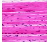
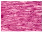
Cardiac Muscle: Involuntary, striated muscle found only in the heart wall. Cells are branched, mostly uninucleated, and connected by intercalated discs containing gap junctions and desmosomes.
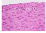
Smooth Muscle: Involuntary, non-striated muscle found in walls of internal organs (e.g., intestines, blood vessels). Cells are spindle-shaped and uninucleated.
Microscopic Anatomy of Skeletal Muscle
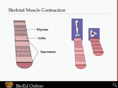
Skeletal muscle fibers are long, cylindrical cells containing multiple nuclei. Each fiber is surrounded by a plasma membrane called the sarcolemma and contains cytoplasm known as sarcoplasm. Within each fiber are myofibrils, which are bundles of actin (thin) and myosin (thick) filaments organized into repeating units called sarcomeres.
Connective Tissue Sheaths
Endomysium: Loose connective tissue surrounding individual muscle fibers.
Perimysium: Dense connective tissue surrounding bundles of fibers (fascicles).
Epimysium: Dense irregular connective tissue surrounding the entire muscle.
Muscle Fiber Structure and Sarcomere Organization
Myofibrils: Rodlike bundles of actin and myosin that run parallel within the muscle cell.
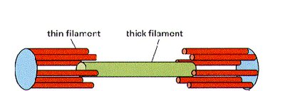
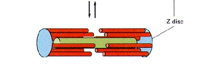
Sarcomere: The functional contractile unit of muscle, defined as the region between two Z discs. The arrangement of actin and myosin gives skeletal muscle its striated appearance.
Key Bands and Lines:
Z disc: Boundary of each sarcomere; anchors thin filaments.
A band: Region containing thick filaments (myosin), with some overlap of thin filaments.
I band: Region containing only thin filaments (actin).
Sliding Filament Theory of Muscle Contraction
Muscle contraction occurs when actin and myosin filaments slide past each other, shortening the sarcomere and thus the muscle fiber. This process is powered by ATP and initiated by a signal from the nervous system.
Motor Units and Neuromuscular Junction
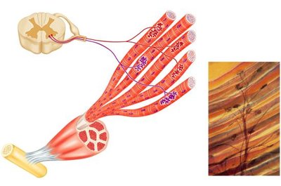
Motor Unit: A single motor neuron and all the muscle fibers it innervates. All fibers in a motor unit contract simultaneously.
Neuromuscular Junction: The synapse where a motor neuron communicates with a muscle fiber to initiate contraction.
Muscle Actions and Coordination
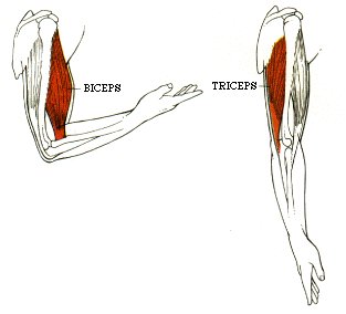
Prime Mover (Agonist): The main muscle responsible for a specific movement.
Antagonist: Muscle that opposes the action of the prime mover.
Synergist: Muscle that assists the prime mover.
Fixator: Muscle that stabilizes the origin of the prime mover.
Muscle Fiber Arrangements
Muscle fibers are organized in different patterns to optimize force, range of motion, or both. Common arrangements include:
Parallel: Fibers run parallel to the muscle's long axis (e.g., sartorius).
Pennate: Fibers attach obliquely to a central tendon (unipennate, bipennate, multipennate; e.g., deltoid).
Convergent: Broad origin with fibers converging to a single tendon (e.g., pectoralis major).
Circular: Fibers arranged in concentric rings (e.g., orbicularis oris).
Summary Table: Comparison of Muscle Tissue Types
Feature | Skeletal Muscle | Cardiac Muscle | Smooth Muscle |
|---|---|---|---|
Control | Voluntary | Involuntary | Involuntary |
Striations | Yes | Yes | No |
Location | Attached to bones | Heart wall | Walls of hollow organs |
Cell Shape | Long, cylindrical, multinucleated | Branched, mostly uninucleated | Spindle-shaped, uninucleated |
Special Features | Motor units, neuromuscular junctions | Intercalated discs, inherent rhythmicity | Regeneration, two layers in organs |
Key Terms and Definitions
Myofilament: Protein filaments (actin and myosin) responsible for muscle contraction.
Sarcolemma: Plasma membrane of a muscle cell.
Sarcomere: The basic contractile unit of muscle fiber.
Motor Unit: A motor neuron and all the muscle fibers it controls.
Neuromuscular Junction: The synapse between a motor neuron and a muscle fiber.
Example: Biceps and Triceps Coordination
During elbow flexion, the biceps brachii acts as the prime mover (agonist), while the triceps brachii serves as the antagonist. Synergists and fixators stabilize the movement, ensuring smooth and efficient action.
Additional info: The protein titin is crucial for muscle elasticity, and the arrangement of muscle fibers within a muscle determines its strength and range of motion. Cardiac muscle's intercalated discs allow for rapid, coordinated contractions essential for heart function.
