 Back
BackMuscle Tissue: Structure, Function, and Physiology
Study Guide - Smart Notes
 Tailored notes based on your materials, expanded with key definitions, examples, and context.
Muscle Tissue Overview
Types of Muscle Tissue
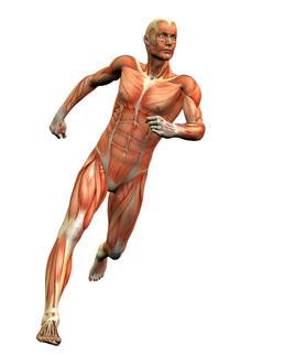
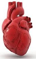
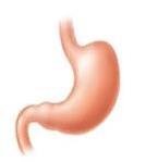
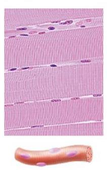
Muscle tissue is specialized for contraction and is essential for movement, posture, and many physiological processes. There are three main types of muscle tissue, each with distinct structure and function:
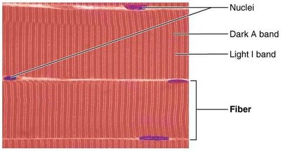
Skeletal Muscle: Attached to bones and skin, responsible for voluntary movements. Skeletal muscle fibers are long, cylindrical, multinucleated, and striated.
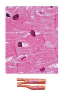
Cardiac Muscle: Found only in the heart, responsible for pumping blood. Cardiac muscle fibers are striated, branched, and interconnected by intercalated discs. Contraction is involuntary.
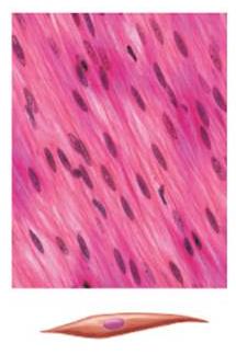
Smooth Muscle: Located in the walls of hollow organs (e.g., stomach, bladder, blood vessels). Smooth muscle fibers are spindle-shaped, non-striated, and contract involuntarily.
Characteristics and Functions of Muscle Tissue
Key Characteristics
Excitability (Responsiveness): Ability to receive and respond to stimuli.
Contractility: Ability to shorten forcibly when stimulated.
Extensibility: Ability to be stretched.
Elasticity: Ability to recoil to resting length after stretching.
Functions
Produce movement (locomotion and manipulation)
Maintain posture and body position
Stabilize joints
Generate heat during contraction
Skeletal Muscle Anatomy
Organization and Connective Tissue Sheaths
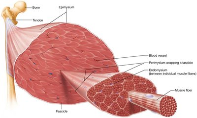
Skeletal muscle is an organ composed of muscle fibers, connective tissue, blood vessels, and nerves. Each muscle and muscle fiber is surrounded by connective tissue:
Epimysium: Surrounds the entire muscle.
Perimysium: Surrounds bundles of muscle fibers (fascicles).
Endomysium: Surrounds each individual muscle fiber.

Muscle Attachments
Direct (fleshy) attachment: Epimysium fused to periosteum or perichondrium.
Indirect attachment: Connective tissue extends as a tendon or aponeurosis.
Muscle Fiber Microanatomy
Structure of a Muscle Fiber
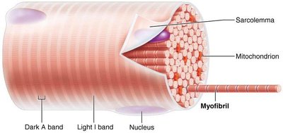
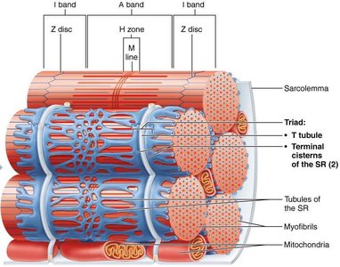
Sarcolemma: Plasma membrane of the muscle fiber.
Sarcoplasm: Cytoplasm containing glycosomes (glycogen storage) and myoglobin (O2 storage).
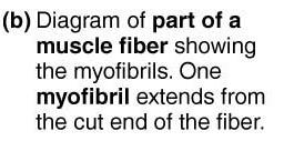
Myofibrils: Densely packed, rodlike elements responsible for muscle contraction and striations.
Sarcoplasmic Reticulum (SR): Specialized endoplasmic reticulum that stores and releases Ca2+.
T Tubules: Invaginations of the sarcolemma that transmit action potentials deep into the fiber.
Myofibrils and Sarcomeres
Striations and Sarcomere Structure
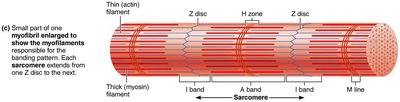
Striations are due to the arrangement of myofilaments (actin and myosin) in repeating units called sarcomeres, the functional unit of muscle contraction.
A bands: Dark regions containing thick (myosin) filaments.
I bands: Light regions containing thin (actin) filaments.
Z disc: Boundary of each sarcomere; anchors thin filaments.
H zone: Lighter region in the middle of the A band.
M line: Line of protein in the center of the H zone.
Myofilament Composition
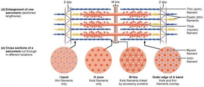
Thick filaments: Composed of myosin molecules with heads that bind to actin during contraction.
Thin filaments: Composed of actin, tropomyosin, and troponin. Actin has binding sites for myosin heads.
Elastic filaments: Composed of titin, which helps maintain sarcomere structure and elasticity.
Muscle Contraction: Sliding Filament Model
Mechanism of Contraction
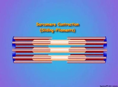
Muscle contraction occurs when thin filaments slide past thick filaments, increasing the overlap and shortening the sarcomere. This process is powered by ATP and regulated by Ca2+ release from the SR.
Myosin heads bind to actin, forming cross bridges.
Cross bridges cycle through attachment, pivoting, detachment, and re-cocking, pulling actin toward the M line.
Sarcomeres shorten, causing muscle contraction.
Neuromuscular Junction and Excitation-Contraction Coupling
Events at the Neuromuscular Junction
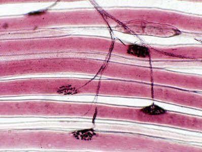
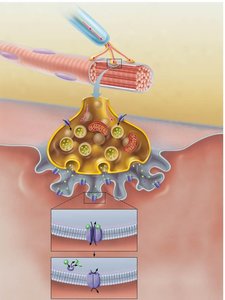
The neuromuscular junction is the site where a motor neuron stimulates a muscle fiber. The process involves:
Action potential arrives at the axon terminal.
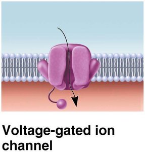
Voltage-gated Ca2+ channels open, Ca2+ enters the neuron.
Ca2+ triggers release of acetylcholine (ACh) into the synaptic cleft.
ACh binds to receptors on the sarcolemma, opening Na+ channels and generating an end plate potential.
ACh is degraded by acetylcholinesterase, terminating the signal.
Muscle Fiber Excitation and Action Potential Generation
End plate potential: Local depolarization due to Na+ influx.
Depolarization: If threshold is reached, voltage-gated Na+ channels open, generating an action potential.
Repolarization: K+ channels open, restoring resting membrane potential.
Excitation-Contraction (E-C) Coupling
E-C coupling links the action potential to muscle contraction:
AP propagates along sarcolemma and down T tubules.
Voltage-sensitive proteins trigger Ca2+ release from SR.
Ca2+ binds to troponin, moving tropomyosin and exposing myosin-binding sites on actin.
Cross bridge cycling begins, leading to contraction.
The Cross Bridge Cycle
Cross bridge formation: Myosin head attaches to actin.
Power stroke: Myosin head pivots, pulling actin toward M line.
Cross bridge detachment: ATP binds to myosin, causing detachment.
Cocking of myosin head: ATP hydrolysis re-energizes the myosin head.
Whole Muscle Contraction
Motor Units and Muscle Twitch
Motor unit: A motor neuron and all the muscle fibers it innervates.
Muscle twitch: Response of a muscle to a single stimulus, consisting of latent, contraction, and relaxation phases.
Graded Muscle Responses
Varying frequency and strength of stimulation produces graded responses.
Wave summation: Increased frequency leads to greater force.
Recruitment: Increasing stimulus strength recruits more motor units.
Types of Contractions
Isotonic contractions: Muscle changes length (concentric or eccentric).
Isometric contractions: Muscle does not change length.
Energy for Contraction
ATP Sources
Direct phosphorylation by creatine phosphate:
Anaerobic glycolysis:
Aerobic respiration:
Muscle Fatigue and Recovery
Fatigue: Inability to contract despite stimulation, often due to ionic imbalances or metabolic byproducts.
Recovery: Oxygen debt (EPOC) must be repaid to restore muscle to pre-exercise state.
Muscle Fiber Types and Adaptation
Classification of Muscle Fibers
Slow oxidative fibers: Endurance, aerobic metabolism.
Fast oxidative fibers: Intermediate, aerobic and anaerobic metabolism.
Fast glycolytic fibers: Short-term, powerful movements, anaerobic metabolism.
Adaptation to Exercise
Aerobic exercise increases capillaries, mitochondria, and myoglobin.
Resistance exercise leads to hypertrophy and increased strength.
Smooth Muscle
Structure and Function
Found in walls of hollow organs, organized in sheets.
Fibers are spindle-shaped, lack striations, and contract involuntarily.
Contraction is slow, sustained, and energy-efficient.
Mechanism of Contraction
Ca2+ enters from extracellular space and SR, binds to calmodulin (not troponin).
Calmodulin activates myosin light chain kinase, which phosphorylates myosin, enabling cross bridge formation.
Types of Smooth Muscle
Unitary (visceral) smooth muscle: Found in most hollow organs, cells connected by gap junctions, contract as a unit.
Multiunit smooth muscle: Found in large airways, arteries, arrector pili, and iris; fibers contract independently.
Comparison of Muscle Types
Feature | Skeletal Muscle | Cardiac Muscle | Smooth Muscle |
|---|---|---|---|
Location | Bones, skin | Heart | Walls of hollow organs |
Striations | Yes | Yes | No |
Control | Voluntary | Involuntary | Involuntary |
Cell Shape | Long, cylindrical, multinucleated | Branched, single nucleus | Spindle-shaped, single nucleus |
Special Features | Fast contraction, tires easily | Intercalated discs, autorhythmic | Gap junctions, slow sustained contraction |
