 Back
Muscle Tissue: Structure, Function, and Physiology
Study Guide - Smart Notes
 Tailored notes based on your materials, expanded with key definitions, examples, and context.
Muscle Tissue Types and Characteristics
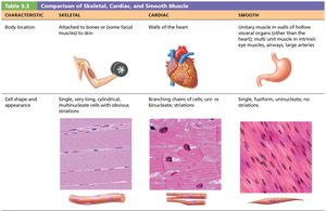
Comparison of Skeletal, Cardiac, and Smooth Muscle
Muscle tissue is classified into three main types: skeletal, cardiac, and smooth. Each type has distinct anatomical and physiological properties that determine its function in the body.
Skeletal Muscle: Attached to bones, responsible for voluntary movements.
Cardiac Muscle: Found only in the heart, responsible for pumping blood.
Smooth Muscle: Located in walls of hollow organs (e.g., intestines, blood vessels), controls involuntary movements.
Key differences:
Cell shape: Skeletal (long, cylindrical, multinucleate), Cardiac (branching, striated, usually one nucleus), Smooth (spindle-shaped, uninucleate, no striations).
Control: Skeletal (voluntary), Cardiac and Smooth (involuntary).
Four Characteristics of Muscle Tissue
Excitability (Responsiveness): Ability to receive and respond to stimuli.
Contractility: Ability to shorten forcibly when stimulated.
Extensibility: Ability to be stretched.
Elasticity: Ability to return to original shape after stretching.
Four Muscle Functions
Movement: Muscles produce movement of body parts and substances.
Posture and Body Position: Muscles maintain posture and stabilize body positions.
Stabilize Joints: Muscles reinforce and stabilize joints.
Generate Heat: Muscle contractions produce heat, helping maintain body temperature.
Skeletal Muscle Anatomy
Structure and Organization of Skeletal Muscle
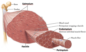
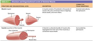
Skeletal muscle is an organ composed of muscle tissue, nerves, blood vessels, and connective tissue sheaths. The organization is hierarchical, from the whole muscle down to individual fibers.
Muscle (organ): Covered by epimysium.
Fascicle: Bundle of muscle fibers, surrounded by perimysium.
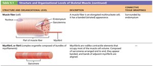
Muscle fiber: Single muscle cell, wrapped by endomysium.
Connective Tissue Sheaths
Endomysium: Surrounds individual muscle fibers.
Perimysium: Surrounds fascicles.
Epimysium: Surrounds the entire muscle.
Muscle Attachments
Muscles attach to bones via two main points:
Origin: Attachment to immovable bone.
Insertion: Attachment to movable bone.
Attachments can be direct (muscle to bone) or indirect (via tendons or aponeuroses).
Neuromuscular Junction and Muscle Contraction
Motor Neurons and Neuromuscular Junction (NMJ)
Skeletal muscle contraction is initiated by signals from motor neurons. The neuromuscular junction is the site where the motor neuron communicates with the muscle fiber.
Motor neuron: Sends action potential down its axon.
Axon terminal: Forms the NMJ with the muscle fiber.
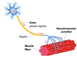
Anatomy of the NMJ
Axon terminal: Contains synaptic vesicles filled with acetylcholine (ACh).
Synaptic cleft: Space between neuron and muscle fiber.
Motor end plate: Region of sarcolemma with ACh receptors.
Junctional folds: Increase surface area for ACh receptors.
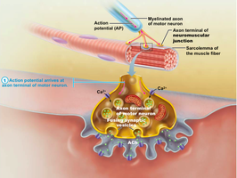
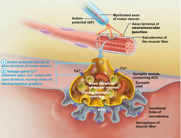
Channels Involved in Initiating Muscle Contraction
Voltage-gated channels: Open in response to changes in membrane potential.
Chemically-gated channels: Open in response to binding of a chemical ligand (e.g., ACh).
Movement of ions (Na+, K+) across the membrane changes the local voltage, leading to muscle contraction.
Action Potential and Muscle Contraction
Depolarization: ACh binding opens Na+ channels, causing influx and depolarization.
Action potential: If depolarization reaches threshold, a wave of voltage-gated Na+ channels opens, spreading the action potential across the sarcolemma.
Repolarization: K+ channels open, restoring resting membrane potential.
Equation for membrane potential:
Muscle Fiber Microanatomy
Muscle Fiber Structure
Sarcolemma: Plasma membrane of muscle fiber.
Sarcoplasm: Cytoplasm, contains glycosomes and myoglobin.
Myofibrils: Contractile protein rods, composed of sarcomeres.
Sarcomere and Myofilaments
Sarcomere: Smallest functional unit, runs from Z disc to Z disc.
Striations: Alternating A bands (dark) and I bands (light).
Myofilaments: Thick (myosin) and thin (actin) filaments.
Excitation-Contraction Coupling
Sarcoplasmic Reticulum and T-Tubules
Sarcoplasmic reticulum (SR): Stores and releases Ca2+.
T-tubules: Invaginations of sarcolemma, bring action potential to SR.
Role of Calcium in Muscle Contraction
Ca2+ binds to troponin, causing a conformational change.
Troponin moves tropomyosin, exposing actin binding sites.
Allows myosin heads to form cross bridges with actin.
Sliding Filament Model and Cross Bridge Cycle
Sliding Filament Model
Myosin heads attach to actin, pull, and detach in a cycle.
ATP is required for detachment and re-cocking of myosin heads.
Cross Bridge Cycle Steps
Cross bridge formation
Power stroke
Cross bridge detachment (bind ATP)
Cocking of myosin head (hydrolyze ATP)
Whole Muscle Contraction
Motor Unit
A motor unit consists of a motor neuron and all the muscle fibers it innervates.
Small motor units allow fine control; large units generate more force.
Muscle Twitch and Graded Contractions
Muscle twitch: Single contraction in response to a single stimulus.
Graded contractions: Vary in strength by changing frequency and recruitment of motor units.
Energy for Muscle Contraction
ATP Sources
Direct phosphorylation: Creatine phosphate regenerates ATP quickly.
Anaerobic pathway: Glycolysis produces ATP and lactic acid.
Aerobic pathway: Glucose and oxygen produce ATP, CO2, and H2O.
Equation for aerobic respiration:
Muscle Fatigue and Recovery
Muscle Fatigue
Inability to contract due to ionic imbalances, depletion of ATP, or accumulation of metabolic byproducts.
Excess Postexercise Oxygen Consumption (EPOC)
Recovery requires replenishing oxygen, converting lactic acid, restoring glycogen, and resynthesizing ATP and CP.
Adaptation and Developmental Aspects
Adaptation to Exercise
Aerobic exercise increases mitochondria, capillaries, and myoglobin.
Resistance exercise increases myofibrils, connective tissue, and muscle size (hypertrophy).
Developmental Aspects
Skeletal muscle regeneration is limited; smooth muscle regenerates well.
Muscle mass differs between sexes due to testosterone.
Aging leads to increased connective tissue and decreased muscle fibers (sarcopenia).
Smooth Muscle
Microscopic Anatomy and Function
Found in walls of hollow organs, lacks striations, has only endomysium.
Cells are spindle-shaped, uninucleate.
Contraction is slow, synchronized, and energy-efficient.
Regulated by neural, hormonal, and local chemical signals.
Special features: Stress-relaxation response, ability to contract over a wide range of lengths.
Additional info: All content is expanded and organized for clarity and completeness, suitable for college-level Anatomy & Physiology study.
