 Back
BackMuscle Tissue: Structure, Function, and Physiology
Study Guide - Smart Notes
 Tailored notes based on your materials, expanded with key definitions, examples, and context.
Muscle Tissue
Types and Functions of Muscle Tissue
Muscle tissue is a primary tissue responsible for contraction and movement in the human body. There are three main types of muscle tissue, each with distinct functions and properties:
Skeletal muscle: Moves the body by pulling on bones; responsible for voluntary movements.
Cardiac muscle: Pumps blood through the cardiovascular system; found only in the heart.
Smooth muscle: Pushes fluids and solids through internal passageways and organs; found in walls of hollow organs.
Common properties of muscle tissue include:
Excitability (responsiveness): Ability to receive and respond to stimuli.
Contractility: Ability of cells to shorten.
Extensibility: Ability of the muscle to stretch.
Elasticity: Ability of the muscle to recoil to its resting length.
Functions of Skeletal Muscle
Skeletal muscles perform several essential functions:
Producing movement: Pull on tendons to move bones.
Maintaining posture and body position.
Supporting soft tissues.
Guarding body entrances and exits: Such as those of the urinary and digestive tract.
Maintaining body temperature: Working muscle produces and releases heat.
Storing nutrients: Source of proteins/amino acids.
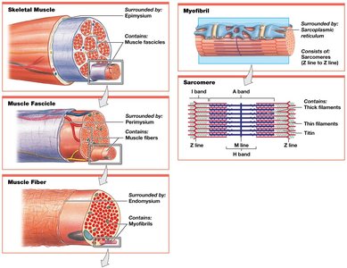
Structure of Skeletal Muscle
Skeletal muscles are composed of muscle tissue, connective tissues, blood vessels, and nerves. They have three layers of connective tissue:
Epimysium: Surrounds the entire muscle; connected to deep fascia.
Perimysium: Surrounds individual fascicles (bundles of muscle fibers).
Endomysium: Surrounds individual muscle cells (muscle fibers).
The collagen fibers of these layers come together at the ends of muscles to form tendons (bundles) or aponeuroses (broad sheets), which attach skeletal muscles to bones.

Blood Supply and Innervation
Skeletal muscles have extensive networks of blood vessels to deliver oxygen and nutrients and remove metabolic wastes.
Muscles contract only when stimulated by the central nervous system; branches of neuronal axons innervate individual muscle fibers.
Skeletal Muscle Fibers
Development and Structure
Skeletal muscle fibers are large, multinucleate cells formed by the fusion of embryonic cells called myoblasts. They are also known as striated muscle cells due to visible striations caused by the arrangement of myofibrils.
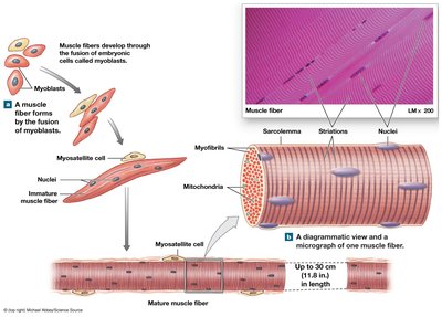
Internal Organization
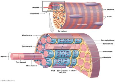
Sarcolemma: Plasma membrane of a muscle fiber; surrounds the sarcoplasm (cytoplasm).
Transverse tubules (T tubules): Narrow tubes continuous with the sarcolemma; transmit action potentials into the cell interior.
Sarcoplasmic reticulum (SR): Tubular network specialized for storage and release of calcium ions; forms terminal cisternae attached to T tubules.
Myofibrils: Organized collections of myofilaments responsible for muscle contraction.
Myofilaments: Bundles of contractile protein filaments; two types: thin (actin) and thick (myosin).
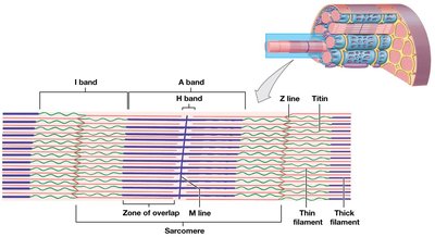
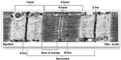
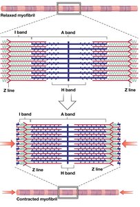
Sarcomere Structure
The sarcomere is the repeating structural and functional unit of a myofibril. It contains thin and thick filaments, proteins that stabilize their positions, and proteins that regulate their interactions.
A bands: Dark bands, length of thick filaments.
M line: Center of A band; stabilizes thick filaments.
H band: Only thick filaments.
Zone of overlap: Region where thick and thin filaments overlap.
I bands: Light bands, only thin filaments.
Z lines: Bisect I bands; mark boundaries between sarcomeres.
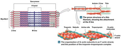
Titin: Elastic protein extending from thick filaments to Z line; maintains alignment and aids in restoring resting length.
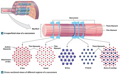
Thin and Thick Filaments
Thin filaments: Composed primarily of actin, with associated proteins (nebulin, tropomyosin, troponin).
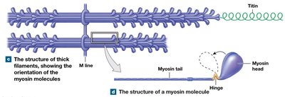
Thick filaments: Composed primarily of myosin; each myosin molecule has a tail (binds to other myosin) and a head (binds to actin).
Titin: Core of thick filaments, acts as a molecular spring.
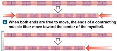
Sliding-Filament Theory
During contraction, thin filaments slide toward the center of the sarcomere (M line) alongside thick filaments. As a result:
H band and I bands narrow
Zones of overlap widen
Z lines move closer together
A band width remains constant
Muscle Contraction
Excitable Membranes and Action Potentials
Muscle cells maintain a negative resting membrane potential. Stimuli can change ion distribution, leading to depolarization, hyperpolarization, or repolarization. Neurons and muscle cells produce action potentials that propagate along the plasma membrane.
Neuromuscular Junction (NMJ)
The synapse between a motor neuron and a skeletal muscle fiber.
Acetylcholine (ACh) is the neurotransmitter released at the NMJ, binding to chemically gated Na+ channels and generating an action potential.
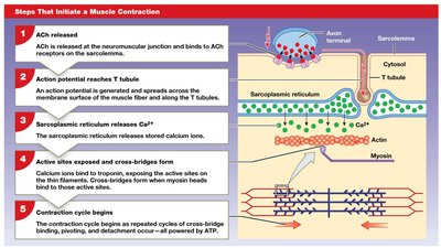
Excitation–Contraction Coupling
The link between the generation of an action potential in the sarcolemma and the start of muscle contraction:
Action potential travels down T tubules to triads.
Triggers release of calcium ions from the SR.
Calcium binds to troponin, moving tropomyosin off actin active sites.
Contraction begins.
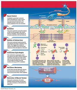
Contraction Cycle
Formation of cross-bridges: Myosin heads bind to actin.
Power stroke: Myosin head pivots, pulling actin toward the M line, using ATP.
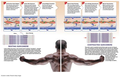
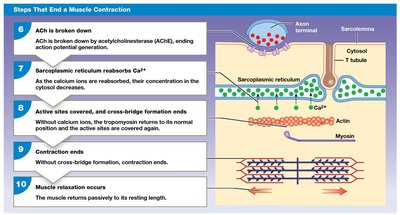
Relaxation
Duration of contraction depends on stimulation, calcium presence, and ATP availability.
When stimulus ends: ACh is broken down, calcium returns to SR, troponin and tropomyosin return to original positions, active sites are covered.
Rigor mortis: Muscle stiffening after death due to ATP depletion and sustained contraction.
Muscle Tension
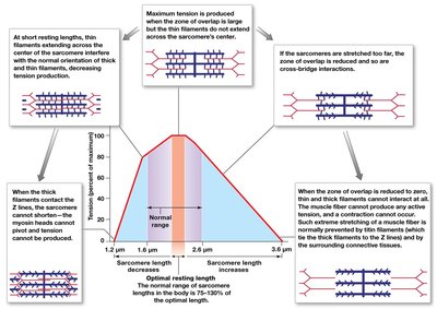
Length–Tension Relationship
The tension produced by a muscle fiber depends on the number of power strokes, resting length, and frequency of stimulation. Maximum tension is produced when the maximum number of cross-bridges is formed at the optimal sarcomere length.
Frequency of Stimulation
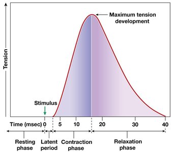
Twitch: Single stimulus-contraction-relaxation sequence.
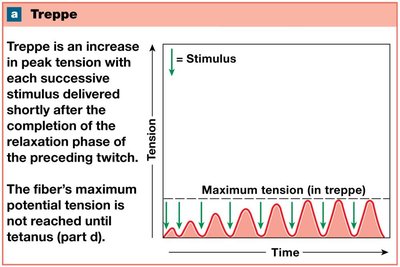
Treppe: Increase in peak tension with repeated stimulations after relaxation phase.
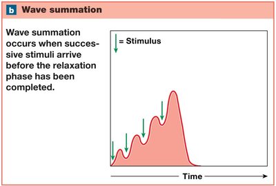
Wave summation: Increasing tension due to successive stimuli before relaxation phase is completed.
Tetanus: Maximum tension; can be incomplete (rapid cycles) or complete (continuous contraction).
Muscle Contractions
Motor Units and Recruitment
Motor unit: A motor neuron and all the muscle fibers it controls.
Recruitment: Increase in muscle tension due to activation of more motor units.
Muscle tone: Resting tension of a skeletal muscle; stabilizes bones and joints.
Types of Muscle Contractions
Isotonic contractions: Muscle changes length.
Isometric contractions: Muscle develops tension but does not change length.
Load and speed of contraction are inversely related.
Return to resting length is a passive process due to elastic forces, opposing muscle contractions, and gravity.
Energy to Power Contractions
ATP Generation and Muscle Fiber Contraction
ATP is the only energy source used directly for muscle contraction.
Muscles store ATP, but stores are depleted quickly; more ATP must be generated.
At rest, ATP transfers energy to creatine to form creatine phosphate (CP).
ATP generation:
Glycolysis: Anaerobic metabolism; net gain of 2 ATP per glucose.
Aerobic metabolism: Oxygen-dependent; produces 15 ATP per pyruvate.
Muscle Metabolism and Activity Levels
At rest: Muscles metabolize fatty acids, store glycogen and CP.
Moderate activity: ATP demand increases; aerobic breakdown of glucose.
Peak activity: ATP demand very high; glycolysis and lactic acid production increase.
Recovery Period and Oxygen Debt
Lactate removal and recycling (Cori cycle): Lactate transferred to liver, converted to pyruvate, then glucose.
Oxygen debt (EPOC): Amount of oxygen required to restore normal conditions after exertion.
Heat production: Muscles release up to 85% of heat needed to maintain body temperature.
Hormones and Muscle Metabolism
Growth hormone and testosterone stimulate protein synthesis and muscle enlargement.
Thyroid hormones elevate energy consumption.
Epinephrine increases muscle metabolism and force of contraction.
Muscle Performance
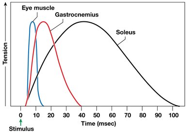
Types of Skeletal Muscle Fibers
Fast fibers: Contract quickly, large diameter, high force, fatigue quickly.
Slow fibers: Contract slowly, small diameter, high endurance, contain myoglobin.
Intermediate fibers: Mid-sized, slower to fatigue than fast fibers.
Most human muscles contain a mixture of fiber types.
Muscle Hypertrophy and Atrophy
Hypertrophy: Muscle growth from heavy training; increase in fiber diameter, myofibrils, mitochondria, and glycogen reserves.
Atrophy: Reduction of muscle size, tone, and power due to lack of activity.
Changes with Aging and Muscle Fatigue
Muscle fibers become smaller, less elastic; tolerance for exercise decreases.
Muscle fatigue: Inability to perform at required level; correlated with depletion of reserves, damage, pH decline, and weariness.
Physical Conditioning
Anaerobic endurance: Supported by stored ATP, CP, and glycolysis; improves muscle power.
Aerobic endurance: Supported by ATP from mitochondria; improves prolonged activity.
Cross-training: Combination of aerobic and anaerobic exercises.
Cardiac Muscle Tissue
Structure and Function
Found only in the heart; small, branched, striated cells with a single nucleus.
Contact via intercalated discs (gap junctions and desmosomes).
Automaticity: Able to contract without neural stimulation; pacemaker cells initiate contraction.
Longer contractions, longer refractory periods, resistance to fatigue.
Contractions require both intracellular and extracellular calcium ions.
No wave summation or tetanic contractions.
Smooth Muscle Tissue
Structure and Function
Found around other tissues and internal organs; spindle-shaped cells with single, central nucleus.
No T tubules, myofibrils, or sarcomeres; nonstriated muscle.
Thin filaments attached to dense bodies; dense bodies connect adjacent cells.
No tendons or aponeuroses.
Functional Characteristics
Excitation–contraction coupling: Calcium ions bind calmodulin, activating myosin light chain kinase.
Length–tension relationships: Plasticity allows function over a wide range of lengths.
Control of contractions: Multiunit and visceral smooth muscle cells.
Smooth muscle tone: Normal background level of activity.
Levers and Muscle Attachments
Levers
Almost all skeletal muscles attach to bones, affecting force, speed, and range of movement. Bones act as levers, joints as fulcrums, and muscles provide the applied force.
First-class lever: Fulcrum between applied force and load (e.g., neck extension).
Second-class lever: Load between applied force and fulcrum (e.g., ankle extension).
Third-class lever: Applied force between load and fulcrum (e.g., elbow flexion).
Origins and Insertions
Origin: Fixed (less movable) point of attachment.
Insertion: More movable point of attachment.
Action: Specific movement produced by muscle contraction.
Muscles work in groups: Agonist (prime mover), antagonist, synergist, fixator.
Muscle opposition: Agonists and antagonists work in pairs; when one contracts, the other stretches or undergoes eccentric contraction.
