 Back
BackMuscle Tissue: Structure, Function, and Physiology
Study Guide - Smart Notes
 Tailored notes based on your materials, expanded with key definitions, examples, and context.
Muscle Tissue Overview
Introduction to Muscle Tissue
Muscle tissue is a specialized tissue responsible for converting chemical energy (ATP) into mechanical movement. It is essential for body motion, posture, and internal transport, comprising about 50% of body mass. Muscle function is closely linked to nervous system control and energy metabolism. Disorders such as muscular dystrophy, spasms, and paralysis highlight the importance of healthy muscle tissue. Muscle cells, known as muscle fibers, are elongated cells containing the contractile proteins actin and myosin that generate force.
Types of Muscle Tissue
Classification and Properties
The human body contains three main types of muscle tissue, each with distinct structure and function:
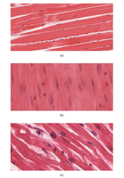
Skeletal Muscle: Voluntary, striated muscle that moves the skeleton.
Cardiac Muscle: Involuntary, striated muscle found only in the heart.
Smooth Muscle: Involuntary, non-striated muscle located in the walls of organs and blood vessels.
All muscle types share four main functional properties:
Excitability: Ability to respond to stimuli (usually from motor neurons).
Contractility: Ability to shorten and generate force.
Extensibility: Ability to stretch without damage.
Elasticity: Ability to return to original length after contraction or stretch.
Skeletal Muscle Structure
Organization and Connective Tissue Layers
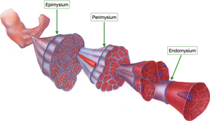
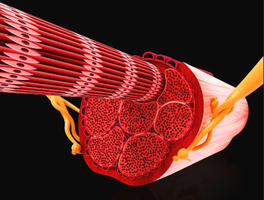
Skeletal muscle is primarily responsible for movement, posture, and heat production, making up about 40% of body weight. It is composed of long, multinucleated muscle fibers organized into bundles called fascicles. These fascicles are surrounded by three layers of connective tissue:
Endomysium: Surrounds individual muscle fibers (innermost layer).
Perimysium: Surrounds fascicles (middle layer).
Epimysium: Surrounds the entire muscle (outermost layer).
Tendons: Dense connective tissue that connects muscle to bone.
These layers protect, support, and organize muscle tissue, facilitate force transmission, and provide pathways for nerves and blood vessels.
Skeletal Muscle Fiber Anatomy
Muscle fibers are elongated, multinucleated cells containing myofibrils, which are responsible for contraction. Myofibrils are composed of repeating units called sarcomeres, which contain actin (thin) and myosin (thick) filaments, giving skeletal muscle its striated appearance. Key structures include:
Sarcolemma: Plasma membrane of the muscle fiber.
T-tubules: Invaginations of the sarcolemma that conduct action potentials deep into the fiber.
Sarcoplasm: Cytoplasm of the muscle fiber.
Sarcoplasmic reticulum: Specialized smooth endoplasmic reticulum that stores and releases calcium ions.
Sarcomere Structure
The sarcomere is the functional unit of muscle contraction, extending from Z-line to Z-line. Its arrangement determines how muscle contracts and generates force. Key regions include:
A-band: Dark region containing thick filaments (myosin).
I-band: Light region containing only thin filaments (actin).
H-zone: Center of the A-band, myosin only.
M-line: Supports myosin filaments.
Z-line (Z-disc): Anchors thin filaments and defines sarcomere boundaries.
Myofilaments and Regulation
Thick filaments are composed of myosin, while thin filaments are made of actin, tropomyosin, and troponin. Tropomyosin and troponin regulate contraction by controlling access to actin's binding sites. Calcium ions bind to troponin, shifting tropomyosin and exposing binding sites for myosin heads to form cross-bridges with actin, initiating contraction.
Muscle Contraction and Relaxation
Cross-Bridge Cycling
The cross-bridge cycle is the process by which myosin heads attach to actin, pivot (power stroke), detach, and reset. Relaxation occurs when calcium ions are pumped back into the sarcoplasmic reticulum, tropomyosin re-covers actin binding sites, and sarcomeres return to resting length. ATP is required for both contraction and relaxation.
Isotonic contraction: Muscle changes length (movement).
Isometric contraction: Muscle length remains the same (tension only).
Nerve Supply and Muscle Activation
Skeletal muscle fibers are activated by motor neurons at the neuromuscular junction (NMJ). The NMJ releases acetylcholine (ACh), triggering an action potential in the muscle fiber, which leads to calcium release and muscle contraction. This process is known as excitation-contraction coupling:
Motor neuron releases ACh at the NMJ.
ACh triggers an action potential in the muscle fiber.
Action potential causes Ca2+ release from the sarcoplasmic reticulum.
Ca2+ binds to troponin, allowing cross-bridge formation and contraction.
Muscle Metabolism and ATP Sources
Blood Supply and Metabolic Support
Skeletal muscle is highly vascularized, with capillaries delivering oxygen, glucose, and fatty acids for ATP production. Blood also removes waste products such as carbon dioxide and lactic acid. Increased capillary density (angiogenesis) enhances endurance in frequently used muscles.
Sources of ATP
ATP is essential for muscle contraction and relaxation. Muscles generate ATP through three main pathways:
Creatine phosphate: Rapid ATP generation for short bursts (~10 seconds).
Glycolysis: Anaerobic breakdown of glucose for moderate bursts (~30-40 seconds).
Aerobic respiration: Mitochondria produce ATP from glucose, fatty acids, and oxygen for sustained activity.
ATP availability limits contraction; metabolic disorders can impair ATP production, leading to weakness and fatigue.
Types of Muscle Contractions
Isotonic and Isometric Contractions
Isotonic contractions: Muscle changes length while producing tension.
Concentric: Muscle shortens (e.g., lifting a weight).
Eccentric: Muscle lengthens while controlling load (e.g., lowering a weight).
Isometric contractions: Muscle produces tension without changing length (e.g., holding a plank).
Nervous System Control of Muscle Tension
Motor Units and Recruitment
A motor unit consists of one motor neuron and all the muscle fibers it activates. The number of fibers per motor unit determines the precision or power of movement:
Fewer fibers: Fine control (e.g., fingers, eyes).
More fibers: High force (e.g., legs, back).
Recruitment is the process of activating more motor units to increase muscle tension.
Stimulation Frequency and Muscle Tone
Twitch: Single stimulus, contraction followed by relaxation.
Wave summation: Second stimulus before full relaxation, increasing force.
Tetanus: Rapid, continuous signals create maximal, sustained contraction.
Treppe: Repeated stimuli after full relaxation, each contraction gets stronger.
Muscle tone: Some motor units fire even at rest; hypertonia (high tone), hypotonia (low tone).
Muscle Fiber Types
Classification by Contraction Speed and Metabolism
Slow Oxidative (SO): Slow contraction, aerobic respiration, high myoglobin and mitochondria, fatigue-resistant.
Fast Oxidative (FO): Fast contraction, primarily aerobic but can use anaerobic glycolysis, intermediate fatigue resistance.
Fast Glycolytic (FG): Fastest contraction, anaerobic glycolysis, low myoglobin and mitochondria, strong force but fatigues quickly.
Exercise and Muscle Performance
Adaptations to Activity
Aerobic (endurance) exercise: Increases mitochondria, capillaries, myoglobin; enhances SO fibers and fatigue resistance.
Resistance (strength) exercise: Increases fiber size (hypertrophy), mainly affects FG fibers, increases force generation.
Disuse: Leads to atrophy, loss of strength, and decreased endurance.
Cardiac Muscle Tissue
Structure and Function
Cardiac muscle tissue is found only in the heart. It is striated and involuntary, with branched fibers connected by intercalated discs that allow rapid electrical communication. Key features include:
Intercalated discs: Contain gap junctions (for electrical signals) and desmosomes (for mechanical strength).
Autorhythmicity: Cardiac fibers can self-generate action potentials without nervous input.
High mitochondria content: Supports continuous aerobic activity.
Smooth Muscle Tissue
Structure and Types
Smooth muscle tissue is found in the walls of hollow organs and blood vessels. It is non-striated, involuntary, and composed of spindle-shaped cells with a single nucleus. Types include:
Single-unit: Fibers connected by gap junctions, contract together for coordinated movement.
Multi-unit: Fibers contract independently, allowing fine control (e.g., iris of the eye).
Key features include calmodulin (binds calcium to trigger contraction) and dense bodies (anchor actin and transmit force).
Function and Special Features
Latch bridges: Sustain tension with minimal ATP, maintaining tone.
Varicosities: Swellings on autonomic axons that release neurotransmitters to control contraction.
Pacesetter cells: Spontaneously depolarize to trigger rhythmic contractions.
Hyperplasia: Smooth muscle can increase cell number, allowing organ growth.
Development and Regeneration of Muscle Tissue
Embryonic Development and Repair
Somites: Blocks of mesoderm in the embryo give rise to muscle tissue.
Myoblasts: Embryonic muscle cells that fuse to form muscle fibers.
Skeletal muscle: Myoblasts fuse to form multinucleated fibers (myotubes).
Cardiac muscle: Myoblasts form branched fibers with intercalated discs.
Smooth muscle: Myoblasts remain single-nucleated.
Regeneration capacity varies:
Skeletal muscle: Limited; satellite cells repair damaged fibers.
Smooth muscle: High regeneration potential; can grow via hypertrophy or hyperplasia, with pericytes assisting in some tissues.
Cardiac muscle: Very limited; injury often replaced by fibrosis.
Summary Table: Types of Muscle Tissue
Feature | Skeletal Muscle | Cardiac Muscle | Smooth Muscle |
|---|---|---|---|
Control | Voluntary | Involuntary | Involuntary |
Striations | Yes | Yes | No |
Location | Skeleton | Heart | Organs, vessels |
Cell Shape | Long, cylindrical, multinucleated | Branched, single nucleus | Spindle-shaped, single nucleus |
Regeneration | Limited (satellite cells) | Very limited (fibrosis) | High (hyperplasia, pericytes) |
Key Formulas and Equations
ATP hydrolysis (energy release):
Creatine phosphate reaction:
Aerobic respiration (simplified):
