 Back
BackLecture 4.2: Muscle Tissue - Structure, Function, and Types
Study Guide - Smart Notes
 Tailored notes based on your materials, expanded with key definitions, examples, and context.
Muscle Tissue Overview
Introduction to Muscle Tissue
Muscle tissue is a primary tissue in the human body, specialized for contraction and movement. There are three main types: skeletal muscle, cardiac muscle, and smooth muscle. Each type has unique structural and functional characteristics, contributing to various physiological roles.
Skeletal muscle: Moves the body by pulling on bones.
Cardiac muscle: Controls the heart's contractions.
Smooth muscle: Regulates movements within internal organs.
Functions of Muscle Tissue
Muscle tissue exhibits several common properties:
Excitability: Ability to respond to stimuli.
Contractility: Ability to shorten and produce force.
Extensibility: Ability to stretch without damage.
Elasticity: Ability to return to original shape after stretching.
Skeletal muscle specifically functions to produce movement, maintain posture, support soft tissues, guard entrances/exits, maintain body temperature, and store nutrients.
Organization of Skeletal Muscle
Structural Hierarchy
Skeletal muscle is organized into several structural levels, each surrounded by connective tissue layers:
Epimysium: Surrounds the entire muscle; connected to deep fascia.
Perimysium: Surrounds bundles of muscle fibers (fascicles); contains blood vessels and nerves.
Endomysium: Surrounds individual muscle fibers; contains capillaries, myosatellite cells, and nerve fibers.
Collagen fibers from these layers converge to form tendons or aponeuroses, which attach muscles to bones.
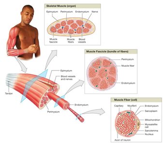
Vascular and Neural Supply
Skeletal muscles have extensive vascular networks for oxygen and nutrient delivery, and waste removal. They contract only when stimulated by the central nervous system, making them voluntary muscles (with exceptions like the diaphragm).
Skeletal Muscle Fibers
Formation and Structure
Skeletal muscle fibers are large, multinucleate cells formed by the fusion of embryonic myoblasts. Myosatellite cells remain as stem cells for repair.
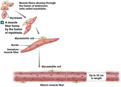
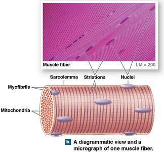
Striated appearance: Due to organized arrangement of myofibrils.
Myofibrils: Cylindrical structures containing myofilaments (actin and myosin).
Sarcolemma: The cell membrane of a muscle fiber.
Sarcomere Structure
Each myofibril contains thousands of sarcomeres, the functional units of contraction. Sarcomeres are composed of overlapping thick (myosin) and thin (actin) filaments, organized into distinct bands:
A band: Both thick and thin filaments; includes H band (thick only) and M line (center).
I band: Thin filaments only; Z line marks boundaries between sarcomeres.
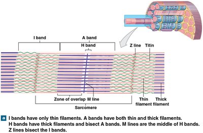
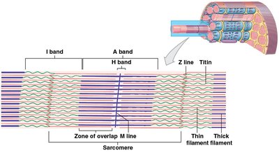
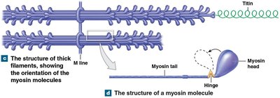
Thick and Thin Filaments
Thick filaments are composed of myosin molecules, while thin filaments are primarily actin. The interaction between these filaments enables muscle contraction.
Mechanism of Muscle Contraction
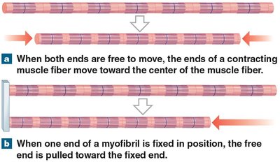
Sliding-Filament Theory
Muscle contraction occurs as thin filaments slide toward the center of the sarcomere, causing the H and I bands to narrow, Z lines to move closer, and the A band to remain constant.
Muscle shortening is an active process; lengthening is passive.
Muscles pull but do not push.
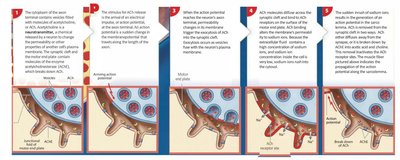
Neuromuscular Junction and Excitation-Contraction Coupling
The neuromuscular junction (NMJ) is the synapse between a motor neuron and a skeletal muscle fiber. The process of muscle contraction involves several steps:
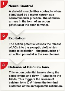
Neural Control: Action potential arrives at axon terminal.
Excitation: Release of acetylcholine (ACh) into synaptic cleft; ACh binds to Na+ channels, depolarizing the motor end plate.
Release of Calcium Ions: Action potential travels along sarcolemma and T tubules, triggering Ca2+ release from sarcoplasmic reticulum.
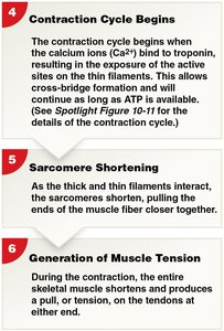
Contraction Cycle Begins: Ca2+ binds to troponin, exposing active sites on actin; cross-bridge formation occurs.
Sarcomere Shortening: Thick and thin filaments interact, shortening sarcomeres.
Generation of Muscle Tension: Muscle produces tension, pulling on tendons.
Types of Muscle Tissue
Cardiac Muscle Tissue
Cardiac muscle cells are found only in the heart. They are striated, have excitable membranes, and are connected by intercalated discs, which allow electrical continuity and coordinated contraction. Cardiac muscle relies on aerobic metabolism and is spontaneously active, with pacemaker cells regulating contraction rate.
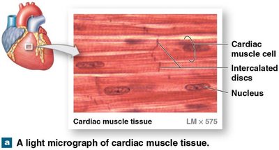
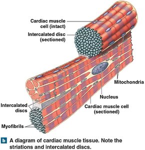
Intercalated discs: Specialized junctions for electrical and mechanical connection.
Spontaneous activity: Cells can contract independently; rate modulated by nervous system.
Sympathetic stimulation: Increases rate and force.
Parasympathetic stimulation: Decreases rate and force.
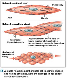
Smooth Muscle Tissue
Smooth muscle is found in sheaths around organs and blood vessels. It lacks striations and sarcomeres, has spindle-shaped cells, and contracts spontaneously. Control is mainly hormonal, with little nervous innervation.
Excitation–contraction coupling: Differs from skeletal and cardiac muscle.
Length–tension relationship: Not directly related; smooth muscle exhibits plasticity.
Control of contractions: Multiunit (innervated) and visceral (pacesetter cells) types.
Smooth muscle tone: Maintains background activity, modulated by neural, hormonal, or chemical factors.
Comparison of Muscle Types
Feature | Skeletal Muscle | Cardiac Muscle | Smooth Muscle |
|---|---|---|---|
Striations | Present | Present | Absent |
Nuclei | Multinucleate | Usually one | One |
Control | Voluntary | Involuntary | Involuntary |
Location | Attached to bones | Heart | Organs, vessels |
Contraction | Rapid, forceful | Rhythmic | Slow, sustained |
Key Terms and Concepts
Myofibril: Cylindrical structure within muscle fiber, composed of myofilaments.
Sarcomere: Functional unit of muscle contraction.
Neuromuscular junction: Synapse between motor neuron and muscle fiber.
Sliding-filament theory: Explains muscle contraction via filament sliding.
Intercalated disc: Specialized junction in cardiac muscle.
Plasticity: Ability of smooth muscle to function over a wide range of lengths.
Important Equations
Muscle force generation is proportional to the number of cross-bridges formed:
Where: = total force = number of cross-bridges = force per cross-bridge
ATP hydrolysis provides energy for contraction:
