 Back
Muscle Tissue: Structure, Organization, and Function
Study Guide - Smart Notes
 Tailored notes based on your materials, expanded with key definitions, examples, and context.
Muscle Tissue
An Introduction to Muscle Tissue
Muscle tissue is a primary tissue type in the human body, specialized for contraction and responsible for producing movement. There are three types of muscle tissue: skeletal muscle, cardiac muscle, and smooth muscle. Each type has unique structural and functional characteristics that enable the body to perform a wide range of activities.
Functions of Muscle Tissue
General Functions
Excitability (Responsiveness): Ability to respond to stimuli.
Contractility: Ability of muscle cells to shorten and generate force.
Extensibility: Ability to be stretched without being damaged.
Elasticity: Ability to return to original length after contraction or extension.
Functions of Skeletal Muscle
Producing movement by pulling on bones
Maintaining posture and body position
Supporting soft tissues
Guarding body entrances and exits
Maintaining body temperature
Storing nutrients
Organization of Skeletal Muscle
Structural Organization
Skeletal muscles are complex organs composed of skeletal muscle tissue, connective tissues, blood vessels, and nerves. The organization of these components allows for efficient force generation and control.
Epimysium: Dense layer of collagen fibers surrounding the entire muscle, separating it from surrounding tissues.
Perimysium: Surrounds bundles of muscle fibers called fascicles; contains blood vessels and nerves.
Endomysium: Surrounds individual muscle fibers; contains capillaries, myosatellite cells (stem cells), and nerve fibers.
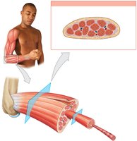
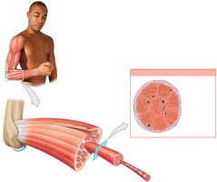
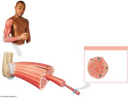
The collagen fibers of all three connective tissue layers merge at the ends of muscles to form tendons (bundles) or aponeuroses (sheets), which attach muscles to bones.
Skeletal Muscle Fibers
Characteristics of Skeletal Muscle Fibers
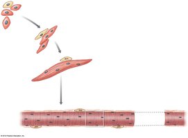
Skeletal muscle fibers are large, multinucleate cells formed by the fusion of embryonic cells called myoblasts. They are also known as striated muscle cells due to their banded appearance.
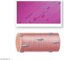
Sarcolemma: The plasma membrane of a muscle fiber, surrounding the sarcoplasm (cytoplasm).
Transverse tubules (T tubules): Invaginations of the sarcolemma that transmit action potentials into the cell interior, triggering contraction.
Sarcoplasmic reticulum (SR): Specialized endoplasmic reticulum that stores and releases calcium ions, essential for muscle contraction.
Myofibrils: Cylindrical structures within muscle fibers, composed of bundles of protein filaments (myofilaments) responsible for contraction.
Levels of Functional Organization
The structure of skeletal muscle is highly organized, from the whole muscle down to the molecular level:
Skeletal muscle (organ) → Muscle fascicle (bundle of fibers) → Muscle fiber (cell) → Myofibril → Sarcomere (functional unit)
Sarcomeres and Myofilaments
Sarcomere Structure
The sarcomere is the smallest functional unit of a muscle fiber. It is defined by the region between two Z lines and is responsible for the striated appearance of skeletal muscle.
A band: Dark region containing thick filaments (myosin) and overlapping thin filaments (actin).
I band: Light region containing only thin filaments.
H band: Central region of the A band with only thick filaments.
M line: Center of the A band; proteins stabilize thick filaments.
Z line: Boundary between adjacent sarcomeres; anchors thin filaments.
Titin: Elastic protein that helps restore sarcomere length after contraction.
Myofilament Composition
Thin filaments: Composed of F-actin (filamentous actin), nebulin, tropomyosin, and troponin. Active sites on G-actin bind to myosin heads during contraction.
Thick filaments: Composed of myosin molecules, each with a tail and two globular heads that interact with actin.
Muscle Contraction: The Sliding Filament Theory
Mechanism of Contraction
According to the sliding filament theory, muscle contraction occurs when thin filaments slide past thick filaments, shortening the sarcomere without changing the length of the filaments themselves. This process is powered by ATP and regulated by calcium ions.
During contraction, H bands and I bands narrow, zones of overlap widen, and Z lines move closer together.
The width of the A band remains constant.
Summary Table: Levels of Skeletal Muscle Organization
Level | Surrounding Structure | Contains |
|---|---|---|
Skeletal Muscle | Epimysium | Muscle fascicles |
Muscle Fascicle | Perimysium | Muscle fibers |
Muscle Fiber | Endomysium | Myofibrils |
Myofibril | Sarcoplasmic reticulum | Sarcomeres |
Sarcomere | — | Thick and thin filaments |
Example: Muscle Fiber Formation
Muscle fibers develop through the fusion of myoblasts, resulting in a multinucleate cell capable of powerful contractions. Myosatellite cells remain associated with muscle fibers and play a role in repair after injury.
Example: Microscopic Structure of a Muscle Fiber
Under the microscope, skeletal muscle fibers display a striated pattern due to the regular arrangement of sarcomeres. Multiple nuclei are located at the periphery of the cell, and the cytoplasm (sarcoplasm) contains numerous myofibrils.
Additional info:
Muscle contraction is initiated by action potentials transmitted from motor neurons to the sarcolemma, leading to calcium release from the sarcoplasmic reticulum and interaction of actin and myosin filaments.
ATP is required for both contraction and relaxation of muscle fibers.
