 Back
Muscles and Compartments of the Lower Appendage
Study Guide - Smart Notes
 Tailored notes based on your materials, expanded with key definitions, examples, and context.
Muscles of the Lower Appendage
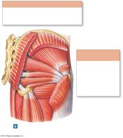
Gluteal and Lateral Rotator Groups
The muscles of the hip and thigh are organized into functional groups based on their actions and anatomical locations. The gluteal group and lateral rotator group are responsible for movements such as extension, abduction, and rotation of the hip.
Gluteus Maximus: Extension and lateral rotation at the hip.
Gluteus Medius & Minimus: Abduction and medial rotation at the hip.
Piriformis: Abduction and lateral rotation at the hip.
Superior/Inferior Gemellus, Obturator Internus, Quadratus Femoris: Lateral rotation at the hip.
Hip Flexors: Iliopsoas Group
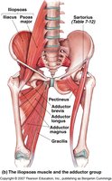
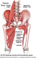
The iliopsoas group is the primary flexor of the hip, composed of the iliacus and psoas major muscles. These muscles are essential for movements such as walking and running.
Iliopsoas: Flexion at the hip.
Muscles That Move the Leg: Sartorius and Gracilis
The sartorius and gracilis muscles contribute to complex movements of the thigh and leg, including flexion, abduction, and rotation.
Sartorius: Flexion, abduction, and lateral rotation of the thigh; helps to cross the leg. Origin: Anterior superior iliac spine. Insertion: Proximal anteromedial aspect of tibia. Innervation: Femoral nerve.
Gracilis: Adduction at the hip, flexion, and medial rotation at the hip.
Quadriceps Femoris Muscle Group
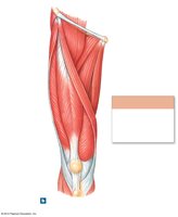
The quadriceps femoris is a powerful muscle group responsible for extension at the knee. It consists of four muscles: rectus femoris, vastus lateralis, vastus medialis, and vastus intermedius.
Rectus Femoris: Extension at the knee. Origin: Anterior inferior iliac spine. Insertion: Tibial tuberosity via quadriceps tendon/patella/patellar ligament. Innervation: Femoral nerve.
Vastus Lateralis, Medialis, Intermedius: Extension at the knee. Origin and insertion vary, but all insert at the tibial tuberosity. Innervation: Femoral nerve.

Adductor Group (Groin Muscles)
The adductor muscles are located in the medial thigh and are responsible for adduction, flexion, and rotation of the hip.
Adductor Brevis/Longus: Adduction, flexion, and medial rotation at the hip.
Adductor Magnus: Adduction; superior part: flexion/medial rotation; inferior part: extension/lateral rotation.
Pectineus and Gracilis: Adduction, flexion, and medial rotation at the hip.
Hamstring Muscle Group
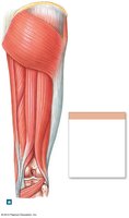
The hamstrings are located in the posterior thigh and are essential for flexion at the knee and extension at the hip.
Biceps Femoris (Long/Short Head): Flexion at the knee. Origin: Ischial tuberosity (long head), linea aspera (short head). Insertion: Head of fibula & lateral condyle of tibia. Innervation: Tibial nerve (long head), common fibular nerve (short head).
Semitendinosus: Flexion at the knee. Origin: Ischial tuberosity. Insertion: Proximal medial surface of tibia. Innervation: Sciatic nerve.
Semimembranosus: Flexion at the knee. Origin: Ischial tuberosity. Insertion: Posterior surface of medial condyle of tibia. Innervation: Tibial/Sciatic nerve.
Muscles That Move the Foot and Toes
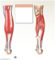
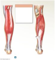
Posterior Compartment: Gastrocnemius, Soleus, Plantaris, Popliteus
These muscles are involved in plantar flexion and stabilization of the ankle and knee.
Gastrocnemius: Plantar flexion at the ankle. Origin: Medial and lateral epicondyles of femur. Insertion: Calcaneus. Innervation: Tibial nerve.
Soleus: Plantar flexion at the ankle. Origin: Fibula, soleal line of tibia. Insertion: Calcaneus. Innervation: Tibial nerve.
Plantaris: Assists in plantar flexion; missing in 10% of people.
Popliteus: Medially rotates tibia on femur (sitting), laterally rotates femur on tibia (standing), unlocks knee for flexion.
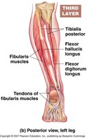
Deep Posterior Compartment: Tibialis Posterior, Flexor Hallucis Longus, Flexor Digitorum Longus
These muscles are responsible for inversion, plantar flexion, and flexion of the toes.
Tibialis Posterior: Inversion of foot and plantar flexion.
Flexor Hallucis Longus: Flexion of the big toe.
Flexor Digitorum Longus: Flexion of toes 2-5.
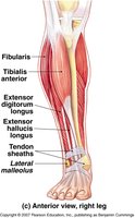
Anterior Compartment: Extensor Hallucis Longus, Extensor Digitorum Longus, Tibialis Anterior
These muscles are involved in dorsiflexion and extension of the toes.
Extensor Hallucis Longus: Extension of the big toe.
Extensor Digitorum Longus: Extension of toes 2-5.
Tibialis Anterior: Dorsiflexion at the ankle.
Lateral Compartment: Fibularis Longus and Brevis
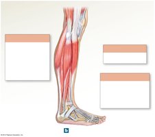
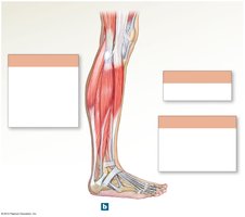
The lateral compartment muscles are responsible for eversion and plantar flexion of the foot.
Fibularis Longus (Peroneus): Eversion of foot and plantar flexion.
Fibularis Brevis: Eversion of foot and plantar flexion.
Intrinsic Muscles of the Foot
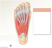
The intrinsic muscles of the foot are responsible for movements of the toes, including flexion and abduction.
Flexor Digitorum Brevis: Flexion of toes 2-5.
Abductor Hallucis: Abduction of the big toe.
Abductor Digiti Minimi: Abduction of the 5th digit.
Fascia, Muscle Layers, and Compartments
Deep Fascia and Musculoskeletal Compartments
Dense connective tissue layers, known as fascia, provide structural support and separate muscles into compartments. These compartments allow for independent movement and contain nerves, blood vessels, and muscle groups.
Deep Fascia: Supports and interconnects adjacent skeletal muscles.
Compartments: Separate soft tissues of the limb into functional groups.


Compartment Syndrome
Compartment syndrome is a medical emergency caused by compression of nerves, blood vessels, and muscle within a closed compartment, leading to tissue death from lack of oxygenation.
Symptoms: Pain, numbness, and loss of function.
Treatment: Surgical intervention to relieve pressure.
