 Back
BackMuscles and Muscle Tissue: Structure and Function
Study Guide - Smart Notes
 Tailored notes based on your materials, expanded with key definitions, examples, and context.
Muscles and Muscle Tissue
Overview of Muscle Tissue
Muscle tissue comprises nearly half of the body’s mass and is specialized for transforming chemical energy (ATP) into mechanical energy, enabling force generation and movement. The study of muscle tissue involves understanding its types, characteristics, and functions.
Types of Muscle Tissue: Skeletal, Cardiac, and Smooth
Key Characteristics: Excitability, Contractility, Extensibility, Elasticity
Main Functions: Movement, posture maintenance, joint stabilization, and heat generation
Types of Muscle Tissue
Skeletal Muscle: Voluntary, striated, attached to bones and skin, responsible for body movement. Skeletal muscle fibers are elongated and called muscle fibers.
Cardiac Muscle: Involuntary, striated, found only in the heart, contracts at a steady rate set by the heart’s pacemaker.
Smooth Muscle: Involuntary, non-striated, found in walls of hollow organs (e.g., stomach, bladder), can contract without nervous stimulation.
Characteristics of Muscle Tissue
Excitability (Responsiveness): Ability to receive and respond to stimuli.
Contractility: Ability to shorten forcibly when stimulated.
Extensibility: Ability to be stretched.
Elasticity: Ability to recoil to resting length after stretching.
Muscle Functions
Produce Movement: Locomotion and manipulation (e.g., walking, digestion, pumping blood).
Maintain Posture and Body Position
Stabilize Joints
Generate Heat: As muscles contract.
Additional Functions: Protect organs, form valves, control pupil size, cause goosebumps.
Skeletal Muscle Anatomy
Structural Organization
Skeletal muscle is an organ composed of muscle tissue, connective tissue, blood vessels, and nerves. It has three main features: nerve and blood supply, connective tissue sheaths, and attachments.
Nerve and Blood Supply: Each muscle receives a nerve, artery, and veins. Nerves control muscle activity, and blood vessels supply nutrients and remove wastes.
Connective Tissue Sheaths: Support and reinforce muscle structure.
Sheath | Location | Description |
|---|---|---|
Epimysium | Surrounds entire muscle | Dense irregular connective tissue |
Perimysium | Surrounds fascicles (muscle fiber bundles) | Fibrous connective tissue |
Endomysium | Surrounds each muscle fiber | Fine areolar connective tissue |
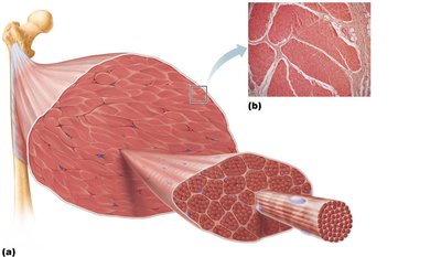
Attachments: Muscles attach to bones at two points: origin (immovable) and insertion (movable). Attachments can be direct (epimysium fused to periosteum) or indirect (via tendons or aponeuroses).
Muscle Fiber Microanatomy and the Sliding Filament Model
Muscle Fiber Structure
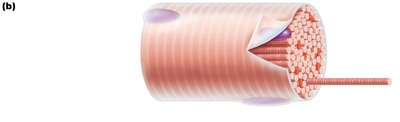
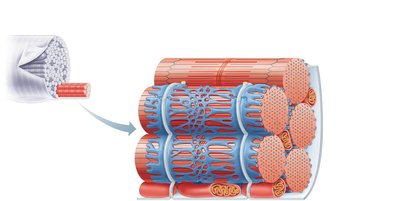
Skeletal muscle fibers are long, cylindrical cells with multiple nuclei. Key components include:
Sarcolemma: Plasma membrane of the muscle fiber
Sarcoplasm: Cytoplasm containing glycosomes (glycogen storage) and myoglobin (O2 storage)
Myofibrils: Densely packed, rodlike elements responsible for muscle contraction
Myofibril Structure
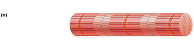
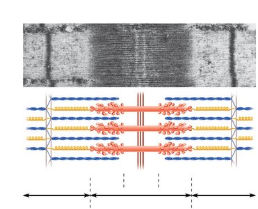
Striations: Alternating dark (A bands) and light (I bands) regions
Sarcomere: The smallest contractile unit, extending from Z disc to Z disc
Myofilaments: Thick (myosin) and thin (actin) filaments arranged in a hexagonal pattern
Molecular Composition of Myofilaments
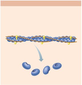
Thick Filaments: Composed of myosin molecules with heads that form cross bridges during contraction
Thin Filaments: Composed of actin, tropomyosin, and troponin; actin has binding sites for myosin heads
Elastic Filaments: Composed of titin, which helps maintain filament alignment and elasticity
Sarcoplasmic Reticulum and T Tubules
Sarcoplasmic Reticulum (SR): Specialized smooth ER that stores and releases Ca2+ for muscle contraction
T Tubules: Invaginations of the sarcolemma that transmit action potentials deep into the muscle fiber
Triad: Structure formed by a T tubule and two terminal cisterns of the SR
Sliding Filament Model of Contraction
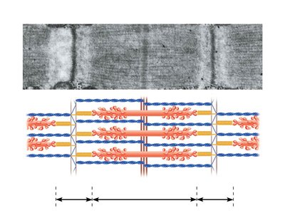
During contraction, thin filaments slide past thick filaments, increasing their overlap. This process is powered by the formation and breaking of cross bridges between actin and myosin heads.
Neither thick nor thin filaments change length; the sarcomere shortens as Z discs are pulled toward the M line.
I bands and H zones shorten, while A bands remain the same length.
Muscle Fiber Contraction
Steps for Skeletal Muscle Contraction
Nerve stimulation
Generation of an action potential in the sarcolemma
Propagation of the action potential along the sarcolemma
Brief rise in intracellular Ca2+ concentration
Steps 1 and 2 occur at the neuromuscular junction; steps 3 and 4 are part of excitation-contraction coupling.
Neuromuscular Junction (NMJ)
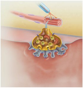
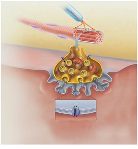
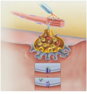
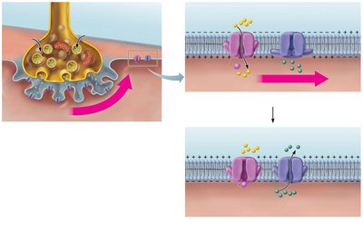
Formed by the axon terminal of a motor neuron and the muscle fiber’s motor end plate, separated by the synaptic cleft.
Acetylcholine (ACh) is released from synaptic vesicles, diffuses across the cleft, and binds to ACh receptors on the sarcolemma, initiating an action potential.
ACh is rapidly broken down by acetylcholinesterase, terminating the signal.
Generation of an Action Potential Across the Sarcolemma
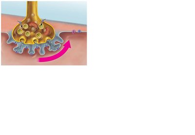
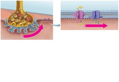
End Plate Potential: Local depolarization due to Na+ influx after ACh binds to its receptor.
Depolarization: If threshold is reached, voltage-gated Na+ channels open, generating an action potential that spreads across the sarcolemma.
Repolarization: Na+ channels close, K+ channels open, restoring the resting membrane potential. The Na+-K+ pump restores ionic conditions.
Excitation-Contraction (E-C) Coupling
E-C coupling links the action potential to muscle contraction. The action potential travels along the sarcolemma and T tubules, triggering Ca2+ release from the SR, which initiates contraction.
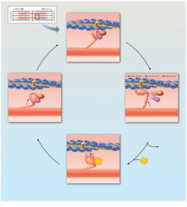
Cross Bridge Cycle
Cross Bridge Formation: Myosin head attaches to actin.
Power Stroke: Myosin head pivots, pulling actin toward the M line.
Cross Bridge Detachment: ATP binds to myosin, causing detachment from actin.
Cocking of Myosin Head: ATP hydrolysis re-energizes the myosin head for the next cycle.
Clinical Note: Rigor Mortis
After death, ATP production ceases, Ca2+ accumulates, and cross bridges form but cannot detach, causing muscle stiffness (rigor mortis) until proteins degrade.
