 Back
BackMuscles and Muscle Tissue: Structure and Function
Study Guide - Smart Notes
 Tailored notes based on your materials, expanded with key definitions, examples, and context.
Muscle Tissue Overview
Types of Muscle Tissue
Muscle tissue is classified into three main types, each with distinct structure, location, and function:
Skeletal Muscle: Attached to bones, responsible for voluntary movements, striated appearance.
Cardiac Muscle: Found only in the heart, involuntary, striated, contracts at a steady rate set by pacemaker cells.
Smooth Muscle: Located in walls of hollow organs (e.g., stomach, blood vessels), involuntary, non-striated, propels substances through internal channels.
Functional Characteristics of Muscle Tissue
All muscle tissues share four key properties:
Excitability (Irritability): Ability to receive and respond to stimuli.
Contractility: Ability to shorten forcibly when stimulated.
Extensibility: Ability to be stretched or extended.
Elasticity: Ability to recoil and resume original length after stretching.
Skeletal Muscle Structure
Organization of Skeletal Muscle
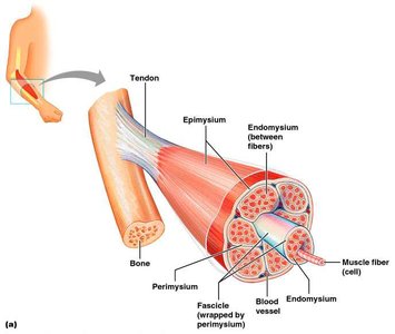
Skeletal muscle is a complex organ composed of muscle fibers, connective tissue, blood vessels, and nerves. The connective tissue sheaths organize and protect muscle fibers at different levels:
Endomysium: Surrounds each individual muscle fiber (cell).
Perimysium: Surrounds groups of muscle fibers, forming fascicles.
Epimysium: Encloses the entire muscle.
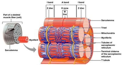
Microscopic Anatomy of a Muscle Fiber
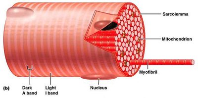
Each skeletal muscle fiber is a long, cylindrical cell with multiple nuclei. Key features include:
Sarcolemma: Plasma membrane of the muscle cell.
Sarcoplasm: Cytoplasm containing glycosomes (glycogen storage) and myoglobin (oxygen-binding protein).
Myofibrils: Densely packed, rodlike contractile elements that make up most of the cell's volume.
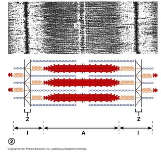
Myofibrils and Sarcomeres
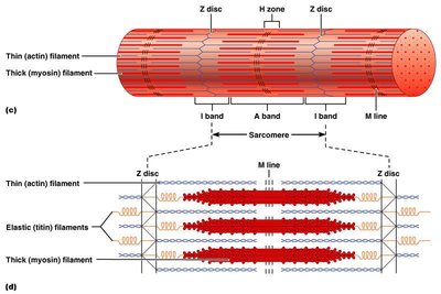
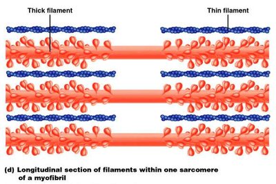
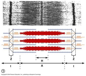
Myofibrils are composed of repeating units called sarcomeres, the smallest contractile units of muscle. Sarcomeres are defined by the region between two Z discs and contain thick (myosin) and thin (actin) filaments arranged in a precise pattern, creating the striated appearance of skeletal muscle.
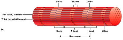
Myofilaments: Thick and Thin Filaments
There are two main types of myofilaments within the sarcomere:
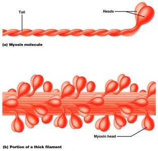
Thick Filaments: Composed of the protein myosin, each molecule has a tail and two globular heads that form cross bridges during contraction.
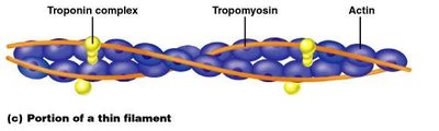
Thin Filaments: Primarily composed of actin, with regulatory proteins tropomyosin and troponin controlling access to myosin-binding sites.
Role of Calcium in Muscle Contraction
Calcium Regulation and the Contraction Mechanism
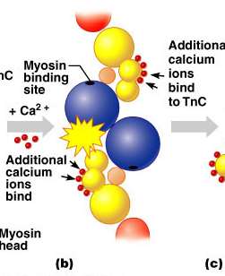
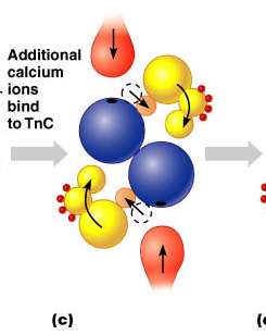
Calcium ions (Ca2+) play a central role in muscle contraction:
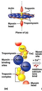
At low Ca2+ concentration, tropomyosin blocks myosin-binding sites on actin, preventing contraction.
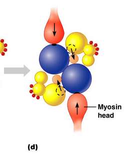
When Ca2+ levels rise, Ca2+ binds to troponin, causing a conformational change that moves tropomyosin away from binding sites, allowing myosin heads to attach and initiate contraction.
Sarcoplasmic Reticulum and T Tubules
Calcium Storage and Release
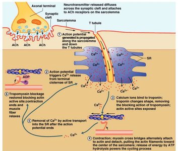
The sarcoplasmic reticulum (SR) is a specialized endoplasmic reticulum that stores and releases Ca2+ in response to electrical signals. T tubules are invaginations of the sarcolemma that conduct action potentials deep into the muscle fiber, ensuring rapid and uniform contraction.

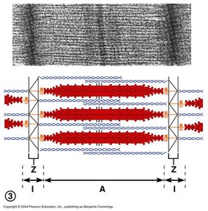
Sliding Filament Model of Contraction
Mechanism of Muscle Shortening
According to the sliding filament model, muscle contraction occurs as thin filaments slide past thick filaments, increasing their overlap and shortening the sarcomere. Myosin heads repeatedly bind to actin, pivot, and pull the thin filaments toward the center of the sarcomere, powered by ATP hydrolysis.
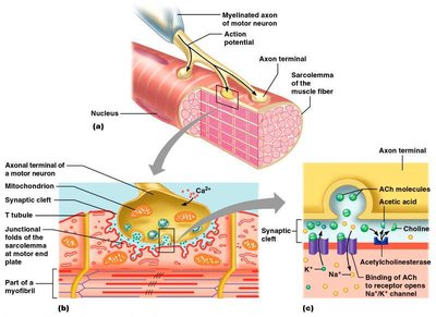
Excitation-Contraction Coupling
From Nerve Signal to Muscle Contraction
Excitation-contraction coupling links the electrical signal from a motor neuron to muscle contraction:
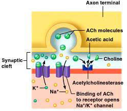
A nerve impulse reaches the neuromuscular junction, causing the release of acetylcholine (ACh).
ACh binds to receptors on the sarcolemma, generating an action potential.
The action potential travels along the sarcolemma and down T tubules, triggering Ca2+ release from the SR.
Ca2+ binds to troponin, exposing myosin-binding sites on actin and initiating contraction.

Contraction Cycle
Cross Bridge Cycling
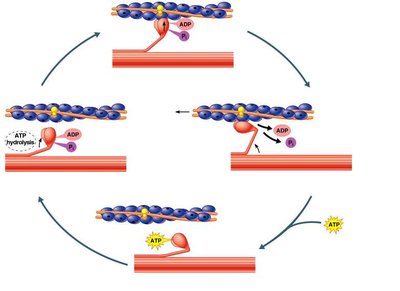
The contraction cycle involves a series of steps:
Myosin head attaches to actin (cross bridge formation).
Power stroke: Myosin head pivots, pulling actin filament toward the M line.
ATP binds to myosin, causing detachment from actin.
ATP is hydrolyzed, re-cocking the myosin head for another cycle.
Motor Units and Muscle Twitch
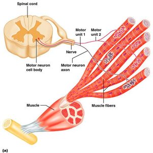
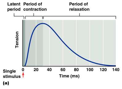
Motor Unit Structure and Function
A motor unit consists of a motor neuron and all the muscle fibers it innervates. The size of a motor unit determines the precision of muscle control. A muscle twitch is the response of a muscle to a single, brief stimulus and consists of three phases: latent period, contraction, and relaxation.
Graded Muscle Responses
Summation and Recruitment
Muscle contractions can be graded by:
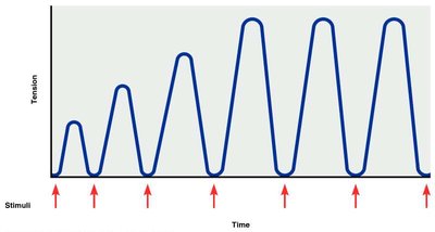
Frequency of stimulation: Increased frequency leads to wave summation and tetanus.
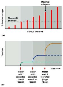
Strength of stimulus: Increased stimulus strength recruits more motor units (recruitment), increasing force.
Treppe (Staircase Effect)
Repeated stimulation of a muscle with the same strength results in progressively stronger contractions due to increased Ca2+ availability and enzyme efficiency.
Types of Muscle Contractions
Isotonic vs. Isometric Contractions
Muscle contractions are classified as:
Isotonic: Muscle changes length and moves a load (e.g., lifting a weight).
Isometric: Muscle develops tension but does not change length (e.g., holding a weight steady).
Muscle Metabolism and Fatigue
Energy Sources for Contraction
ATP is the direct energy source for muscle contraction. It is regenerated by:
Creatine phosphate (CP) pathway
Anaerobic glycolysis (produces lactic acid)
Aerobic respiration
Muscle Fatigue and Oxygen Debt
Muscle fatigue occurs when ATP production cannot keep up with demand, lactic acid accumulates, and ionic imbalances develop. After exercise, oxygen debt must be repaid to restore muscle to its resting state.
Smooth Muscle Structure and Function
Organization and Contraction
Smooth muscle fibers are spindle-shaped, non-striated, and found in the walls of hollow organs. They contract via the sliding filament mechanism, but lack sarcomeres and have different regulatory proteins (calmodulin instead of troponin).
Types of Smooth Muscle
Single-unit (visceral): Cells are electrically coupled and contract as a unit.
Multiunit: Cells function independently, found in large airways, arteries, and eye muscles.
Muscular Dystrophy
Definition and Effects
Muscular dystrophy refers to a group of inherited diseases characterized by progressive muscle weakness and degeneration, often due to abnormal muscle proteins.
