 Back
Muscles and Muscle Tissue: Structure and Function
Study Guide - Smart Notes
 Tailored notes based on your materials, expanded with key definitions, examples, and context.
Muscle Tissue Types and Characteristics
Types of Muscle Tissue
Muscle tissue is classified into three main types, each with distinct structural and functional properties:
Skeletal Muscle: Voluntary muscle responsible for body movement. Contracts rapidly, tires easily, and is powerful.
Cardiac Muscle: Involuntary muscle found only in the heart. Specialized for rhythmic contraction.
Smooth Muscle: Involuntary muscle found in walls of hollow organs (e.g., intestines, blood vessels).
Characteristics of Muscle Tissue
All muscle tissues share four fundamental characteristics:
Excitability (Responsiveness): Ability to receive and respond to stimuli.
Contractility: Ability to shorten forcibly when stimulated.
Extensibility: Ability to be stretched beyond resting length.
Elasticity: Ability to recoil to original length after stretching.
Functions of Muscle Tissue
Muscle tissue performs several essential functions:
Produce Movement: Enables locomotion, digestion, and circulation.
Maintain Posture and Body Position: Stabilizes the body during activity.
Stabilize Joints: Reinforces and supports joints.
Generate Heat: Muscle contraction produces heat, helping maintain body temperature.
Macroscopic Anatomy of Skeletal Muscle
Connective Tissue Sheaths
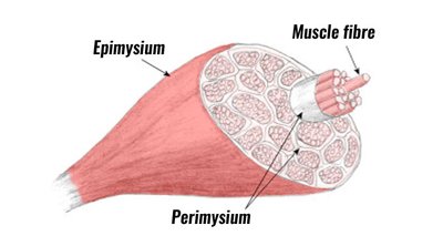
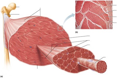
Skeletal muscle is organized into layers of connective tissue sheaths:
Epimysium: Surrounds the entire muscle.
Perimysium: Surrounds groups of muscle fibers called fascicles.
Endomysium: Surrounds individual muscle fibers.
Nerve and Blood Supply
Each muscle fiber is supplied by nerves and blood vessels:
Nerves: Control muscle activity.
Arteries and Veins: Deliver oxygen and nutrients, remove waste products.
Muscle Attachments
Muscles attach to bones at two main points:
Insertion: Attachment to movable bone.
Origin: Attachment to immovable or less movable bone.
Attachments can be:
Direct: Epimysium fused to periosteum of bone.
Indirect: Connective tissue extends as a tendon or aponeurosis.
Microscopic Anatomy of Skeletal Muscle
Muscle Fiber Structure
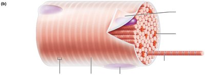
Skeletal muscle fibers are long, cylindrical cells with multiple nuclei. Key components include:
Sarcolemma: Plasma membrane of the muscle fiber.
Sarcoplasm: Cytoplasm containing glycosomes (glycogen storage) and myoglobin (oxygen storage).
Myofibrils and Striations
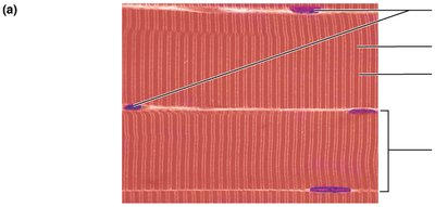
Myofibrils are rod-like organelles that make up most of the muscle cell volume. They display striations due to the arrangement of:
A bands: Dark regions.
I bands: Light regions.
H zone: Central lighter region within A band.
M line: Middle of H zone.
Z disc: Boundary of each sarcomere.
Myofibrils (organelle) = densely packed
accounts for 80% of muscle cell volume
Sarcomere: Functional Unit
The sarcomere is the smallest contractile unit of muscle fiber, extending from one Z disc to the next. Sarcomeres align end-to-end along myofibrils.
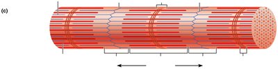
Myofilaments
Myofilaments are the contractile proteins within the sarcomere:
Actin (Thin) Filaments: Extend across I band and partway into A band; anchored to Z discs.
Myosin (Thick) Filaments: Extend the length of A band; connected at M line.
Elastic (Titin) Filaments: Help maintain organization and elasticity.
Myosin and Actin Structure
Myosin: Each thick filament consists of many myosin molecules with protruding heads that bind actin and ATP.
Actin: Thin filaments are composed of two strands of actin subunits twisted into a helix, plus regulatory proteins (troponin and tropomyosin).
G actin subunits link together to form , Fibrous F -actin
Two F actin strands twist together to form a thin filament
Tropomyosin and troponin - regulatory proteins bound to actin
Myosinbinding sites
Troponin: Structure and Function in Muscle Contraction
Troponin is a globular protein found in skeletal and cardiac muscle, and it plays a crucial regulatory role in muscle contraction.
It is part of the thin filament (actin) complex within the sarcomere, the fundamental unit of muscle contraction.
🧬 Location: Troponin is attached to both actin (the thin filament) and tropomyosin (a thread-like protein that wraps around actin).
🔒 Function: At rest, tropomyosin blocks the myosin binding sites on actin, preventing contraction. Troponin acts as a "gatekeeper" for these sites.
⚡ Calcium Activation: When a muscle receives a signal to contract, calcium ions are released from the sarcoplasmic reticulum. Calcium binds to troponin, causing it to change shape.
🔓 Unblocking the Binding Site: This shape change pulls tropomyosin away from the myosin binding sites on actin, allowing myosin to bind and initiate contraction.
💡 Summary: Troponin enables muscle contraction by responding to calcium and moving tropomyosin, so myosin can pull on actin and shorten the sarcomere.
Memory Tip: Tropomyosin says "no" to myosin (blocks contraction), while troponin says "go" (opens the binding site when calcium is present). If you want to learn more about the step-by-step process of muscle contraction or the roles of other proteins like myosin, actin, or titin, just ask!
Sarcoplasmic Reticulum and T Tubules
Sarcoplasmic Reticulum (SR): Specialized smooth endoplasmic reticulum surrounding each myofibril; stores and releases Ca2+.
T Tubules: Invaginations of the sarcolemma that conduct action potentials into the muscle fiber.
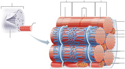
Skeletal Muscle Contraction
Sliding Filament Model
Muscle contraction occurs when thin filaments slide past thick filaments, increasing overlap. Neither filament changes length; instead, the sarcomere shortens.
Cross bridges: Form and break repeatedly, pulling Z discs toward M line.
Band changes: I bands shorten, H zones disappear, A bands move closer.
Regulation of Muscle Fiber Contraction
Contraction is regulated by neural signals:
Motor Neurons: Activate muscle fibers via neuromuscular junction (NMJ).
Acetylcholine (ACh): Neurotransmitter that triggers muscle action potential.
Action potentials = Muscle and nerve cells are the only cell that are excitable
Neuromuscular Junction (NMJ)
The NMJ is the site where motor neuron axon terminals meet the muscle fiber. Key steps:
Action potential arrives at axon terminal.
Voltage-gated Ca2+ channels open; Ca2+ enters axon terminal.
ACh released by exocytosis.
ACh diffuses across synaptic cleft and binds to receptors on sarcolemma.
Chemically gated ion channels open; Na+ enters, K+ exits, producing end plate potential (EPP).
ACh effects terminated by acetylcholinesterase.
Muscle Fiber Excitation
Generation of an action potential across the sarcolemma involves:
End Plate Potential: Local depolarization from Na+ influx.
Depolarization: Voltage-gated Na+ channels open, AP propagates.
Repolarization: Na+ channels close, K+ channels open, restoring resting potential.
Excitation-Contraction Coupling
Excitation-contraction coupling links the action potential to muscle contraction:
AP propagates along sarcolemma and down T tubules.
Ca2+ released from SR.
Ca2+ binds to troponin, shifting tropomyosin and exposing myosin-binding sites.
Contraction begins as myosin binds actin (cross bridge cycling).
Cross Bridge Cycling
Cross bridge cycling is the process by which myosin heads pull actin filaments:
Cross Bridge Formation: Myosin head binds to actin.
Power Stroke: Myosin head pivots, pulling actin toward M line.
Detachment: ATP binds to myosin, causing detachment from actin.
Cocking: ATP hydrolysis re-energizes myosin head.
The Big Picture: Skeletal Muscle Contraction (4-Step Overview)
You're right! The process is often summarized in four main steps, especially when focusing on the essentials at the cellular level. Here’s the streamlined "big picture" for skeletal muscle contraction:
1. Neural Signal & Neuromuscular Junction 🧠 A motor neuron releases acetylcholine at the neuromuscular junction, triggering an action potential in the muscle fiber’s membrane (sarcolemma).
2. Excitation-Contraction Coupling ⚡ The action potential travels along the sarcolemma and dives into T-tubules, stimulating the sarcoplasmic reticulum to release calcium ions into the sarcomere.
3. Calcium & Regulatory Proteins 🧪 Calcium binds to troponin, causing tropomyosin to move and expose myosin-binding sites on actin.
4. Cross-Bridge Cycle & Contraction 💪 Myosin heads bind to actin, perform a power stroke (pulling actin), then release and reset using ATP. This cycle repeats, shortening the sarcomere and contracting the muscle.This cycle repeats as long as ATP and Ca2+ are available.
Rigor Mortis
After death, ATP production ceases, Ca2+ cannot be pumped back into SR, and cross bridge detachment does not occur, resulting in sustained muscle contraction (rigor mortis).
Summary Table: Muscle Tissue Types
Type | Location | Control | Function |
|---|---|---|---|
Skeletal | Attached to bones | Voluntary | Movement, posture |
Cardiac | Heart | Involuntary | Pumping blood |
Smooth | Walls of hollow organs | Involuntary | Movement of substances |
Key Equations
Resting Membrane Potential:
ATP Hydrolysis:
Additional info:
These notes expand upon the original slides by providing definitions, examples, and context for each topic, ensuring a comprehensive and self-contained study guide for exam preparation.
