 Back
Muscles and Muscle Tissue: Structure, Function, and Electromyography
Study Guide - Smart Notes
 Tailored notes based on your materials, expanded with key definitions, examples, and context.
Muscle Tissue Types
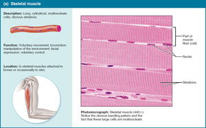
Skeletal Muscle
Skeletal muscle is one of the three main types of muscle tissue, characterized by its voluntary control and striated appearance. It is responsible for movement, posture, and facial expressions.
Microscopic Anatomy: Long, cylindrical, multinucleate cells with obvious striations.
Location: Attached to bones via tendons.
Function: Voluntary movement, locomotion, manipulation of the environment.
Fatigability: Contracts quickly and tires easily.
Skeletal Muscle Cells
Long, cylindrical, striated, and multinucleate.
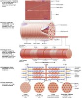
Striations are due to the arrangement of contractile proteins (actin and myosin) in sarcomeres.
Sarcomeres
Functional unit of striated muscle contraction.
Bounded by Z discs; contains A bands (thick and thin filaments overlap), I bands (thin filaments only), H zone (thick filaments only), and M line (anchors thick filaments).
Sliding filament mechanism: contraction occurs as actin and myosin filaments slide past each other.
Cardiac Muscle
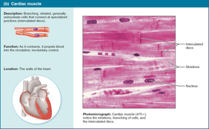
Cardiac muscle forms the myocardium of the heart and is responsible for pumping blood throughout the body. It is involuntary and exhibits autorhythmicity.
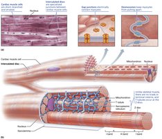
Microscopic Anatomy: Striated, branched, uninucleate cells with intercalated discs.
Location: Heart wall.
Function: Involuntary contraction to propel blood.
Fatigability: Non-fatigable, fast contraction.
Intercalated discs: Specialized junctions containing gap junctions and desmosomes for electrical and mechanical connectivity.
Smooth Muscle
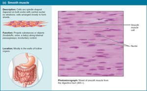
Smooth muscle is found in the walls of hollow organs and is responsible for involuntary movements such as peristalsis and vasoconstriction.
Microscopic Anatomy: Spindle-shaped, uninucleate, non-striated cells.
Location: Digestive, respiratory, reproductive tracts, blood vessels, arrector pili, pupil.
Function: Propels substances, regulates internal movements.
Fatigability: Long, slow contractions, non-fatigable.
Organization: Actin and myosin are not organized into sarcomeres; fibers attach to dense bodies.
Muscle Functional Groups
Classification by Function
Muscles are grouped based on their roles in movement.
Prime mover (agonist): Main muscle responsible for movement.
Antagonist: Opposes or reverses the movement.
Synergist: Assists the prime mover.
Fixator: Immobilizes the origin of the prime mover.
Naming Skeletal Muscles
Conventions in Muscle Naming
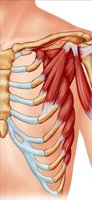
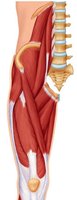
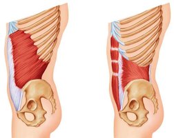
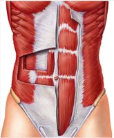
Muscles are named based on several criteria:
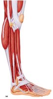
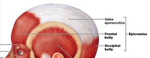
Location: e.g., Epicranius (around cranium), Tibialis anterior (front of tibia).
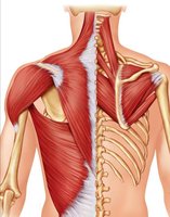
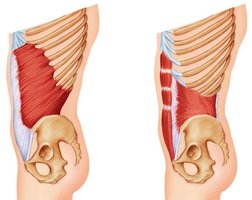
Shape: e.g., Deltoid (triangle), Trapezius (trapezoid), Serratus (saw-toothed), Rhomboideus (rhomboid), Orbicularis (circular).
Size: e.g., Maximus (largest), Minimus (smallest), Longus (longest), Brevis (short), Major (large), Minor (small).
Direction of fibers: e.g., Rectus (straight), Oblique (angled), Transverse (right angles).
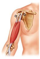
Number of origins: e.g., Biceps (2), Triceps (3), Quadriceps (4).
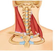
Origin and insertion: e.g., Sternocleidomastoid (originates from sternum and clavicle, inserts on mastoid process).
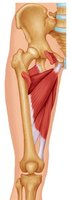
Action: e.g., Flexor carpi radialis (flexes wrist), Abductor pollicis brevis (abducts thumb), Adductor magnus (adducts thigh), Extensor digitorum (extends fingers).
Connective Tissue Coverings of Skeletal Muscle
Organization and Function
Skeletal muscle is organized into layers of connective tissue that provide structure and support.
Superficial fascia: Hypodermis, separates muscle from skin.
Deep fascia: Lines body walls, binds muscles into groups.
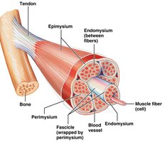
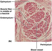
Epimysium: Wraps entire muscle.
Perimysium: Surrounds fascicles (bundles of muscle fibers).
Endomysium: Wraps individual muscle fibers.
Attachments to Bone
Origin: Attachment to stationary bone.
Insertion: Attachment to bone that moves.
Direct attachment: Connective tissue fused to periosteum.
Indirect attachment: Connective tissue forms tendon (cord) or aponeurosis (sheet).
Neuromuscular Junction
Structure and Function
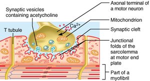
The neuromuscular junction is the site where a motor neuron communicates with a skeletal muscle fiber, initiating contraction.
Axon terminal: Releases neurotransmitter (acetylcholine).
Motor end plate: Specialized region of muscle membrane.
Synaptic cleft: Gap between neuron and muscle.
Muscle Contraction and Electromyography
Motor Unit and Myogram
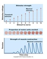
Motor unit: Motor neuron and all muscle fibers it innervates.
Myogram: Recording of muscle contraction.
All-or-none rule: All fibers in a motor unit contract fully.
Twitch: Single contraction from one stimulus.
Phases of Myogram
Latent period: Delay between stimulus and contraction.
Contraction phase: Muscle shortens or develops tension.
Relaxation phase: Muscle returns to resting length.
Refractory period: Muscle cannot respond to new stimulus.
Graded Muscle Responses
Twitch: Single contraction.
Wave (temporal) summation: Repeated contractions before relaxation, leading to stronger contractions.
Treppe: Staircase effect, muscle "warms up" with repeated stimulation.
Multiple Motor Unit Recruitment (MMUR): More motor units recruited for greater force.
Types of Muscle Contraction
Isometric: Muscle does not shorten, tension increases.
Isotonic: Muscle shortens, tension remains constant.
Force of Contraction
Number of fibers contracting (recruitment).
Size of muscle.
Series elastic elements.
Degree of muscle stretch.
Electromyography (EMG) and Biopac Exercise
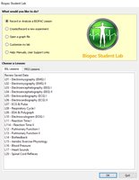
EMG II: Biopac Lesson 2
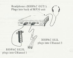
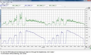
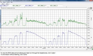
Electromyography (EMG) is used to record electrical activity in muscles. The Biopac system allows students to measure muscle contraction and fatigue.
Attach electrodes to arms.
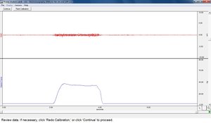
Calibrate dynamometer for zero and maximum force.
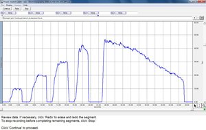
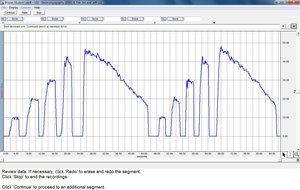
Record clench force at 25%, 50%, 75%, and 100% of maximum.
Observe muscle fatigue by maintaining maximal clench until force drops by 50%.
Data Analysis
Highlight plateaus for MMUR (Multiple Motor Unit Recruitment) on force graph.
Measure fatigue by recording force from maximum to 50% of maximum.
Print and label graphs for lab report.
Key Terms and Concepts
Myology: Scientific study of muscle.
Muscle fiber: Muscle cell.
Myo, mys, sarco: Roots referring to muscle.
Prime mover, antagonist, synergist, fixator: Functional muscle groups.
Origin, insertion: Muscle attachment points.
Motor unit: Motor neuron and connected muscle fibers.
Electromyography (EMG): Recording of muscle electrical activity.
Formulas and Calculations
Percentage of Maximal Clench Force
To calculate the force needed for a given percentage of maximal clench force:
Let Fmax be the maximal clench force.
Force at x%:
Example: If Fmax = 48 kg, then 25% is kg.
Summary Table: Muscle Tissue Types
Type | Microscopic Anatomy | Location | Control | Fatigability |
|---|---|---|---|---|
Skeletal | Striated, multinucleate, cylindrical | Attached to bones | Voluntary | Fatigable |
Cardiac | Striated, branched, uninucleate, intercalated discs | Heart wall | Involuntary | Non-fatigable |
Smooth | Nonstriated, spindle-shaped, uninucleate | Walls of hollow organs | Involuntary | Non-fatigable |
Example: Muscle Naming
Deltoid: Named for its triangular shape.
Biceps brachii: Named for two origins.
Sternocleidomastoid: Named for origin (sternum, clavicle) and insertion (mastoid process).
Flexor carpi radialis: Named for action (flexes wrist).
Additional info:
Muscle fatigue is a decrease in the ability to generate force, often due to depletion of energy stores or accumulation of metabolic byproducts.
Electromyography is a valuable tool for studying muscle physiology and recruitment patterns.
